

 PDF
PDF ePub
ePub Citation
 Print
Print


Abstract
Purpose
To report a case of choroidal neovascularization (CNV) secondary to candida chorioretinitis initially treated with an intravitreal bevacizumab injection.
Case summary
A 50-year-old female presented at our clinic with decreased vision and metamorphopsia in her left eye of 5 days duration. She received an anti-fungal treatment 2 months prior due to the presence of endogenous candida choroiditis in both eyes. Fluorescein angiography and optical coherence tomography (OCT) revealed juxtafoveal CNV in her left eye. Three monthly intravitreal injections of bevacizumab were administered as the initial loading dosage. Her visual symptoms improved and CNV regression was observed on OCT. No recurrence or complications were observed during the 6 month follow-up.
References
1. Jampol LM, Sung J, Walker JD, et al. Choroidal neovascularization secondary to Candida albicans chorioretinitis. Am J Ophthalmol. 1996; 121:643–9.

2. Beebe WE, Kirkland C, Price J. A subretinal neovascular membrane as a complication of endogenous Candida endophthalmitis. Ann Ophthalmol. 1987; 19:207–9.
3. Tedeschi M, Varano M, Schiano Lomoriello D, et al. Photodynamic therapy outcomes in a case of macular choroidal neovascularization secondary to Candida endophthalmitis. Eur J Ophthalmol. 2007; 17:124–7.
4. CATT Research Group. Ranibizumab and bevacizumab for neo- vascular age-related macular degeneration. N Engl J Med. 2011; 364:1897–908.
5. Ruiz-Moreno JM, Montero JA. Intravitreal bevacizumab to treat myopic choroidal neovascularization: 2-year outcome. Graefes Arch Clin Exp Ophthalmol. 2010; 248:937–41.
6. Julian K, Terrada C, Fardeau C, et al. Intravitreal bevacizumab as first local treatment for uveitis-related choroidal neovascularization: long-term results. Acta Ophthalmol. 2011; 89:179–84.
7. Ehrlich R, Ciulla TA, Maturi R, et al. Intravitreal bevacizumab for choroidal neovascularization secondary to presumed ocular histoplasmosis syndrome. Retina. 2009; 29:1418–23.
8. Schadlu R, Blinder KJ, Shah GK, et al. Intravitreal bevacizumab for choroidal neovascularization in ocular histoplasmosis. Am J Ophthalmol. 2008; 145:875–8.
9. Lalwani GA, Rosenfeld PJ, Fung AE, et al. A variable-dosing regimen with intravitreal ranibizumab for neovascular age-related macular degeneration: year 2 of the PrONTO Study. Am J Ophthalmol. 2009; 148:43–58.e1.
10. Sheu SJ. Intravitreal ranibizumab for the treatment of choroidal neovascularization secondary to endogenous endophthalmitis. Kaohsiung J Med Sci. 2009; 25:617–21.
Figure 1.
(A) A colored fundus photograph of a patient with endogenous Candida chorioretinitis showing multifocal yellow-white chorioretinal infiltrates in both the eyes. (B) Color fundus photographs after systemic anti-fungal therapy. A reduced number of multiple yellow-white infiltrates and some chorioretinal scars remained in both eyes. (C, D, E) Imaging studies of the patient who had a visual disturbance in the left eye two months after anti-fungal therapy administration. (C) Color fundus photograph showing subretinal hemorrhage and subretinal membrane temporal to the left fovea. (D) Fluorescein angiography demonstrates a late juxtafoveal leakage. (E) Subretinal fluid and a subretinal neovascular complex are visible on the spectral-domain optical coherence tomography image.
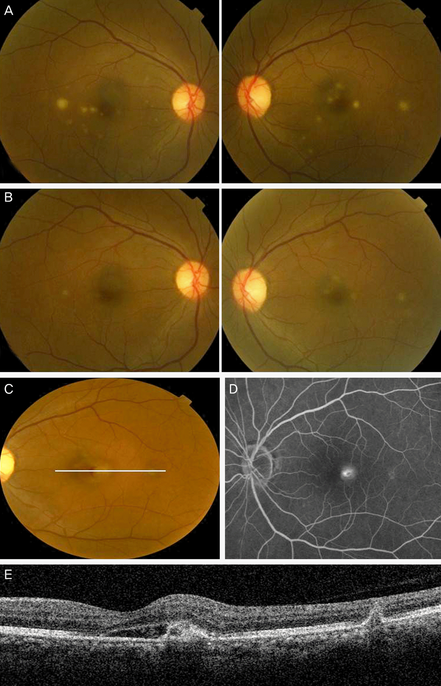
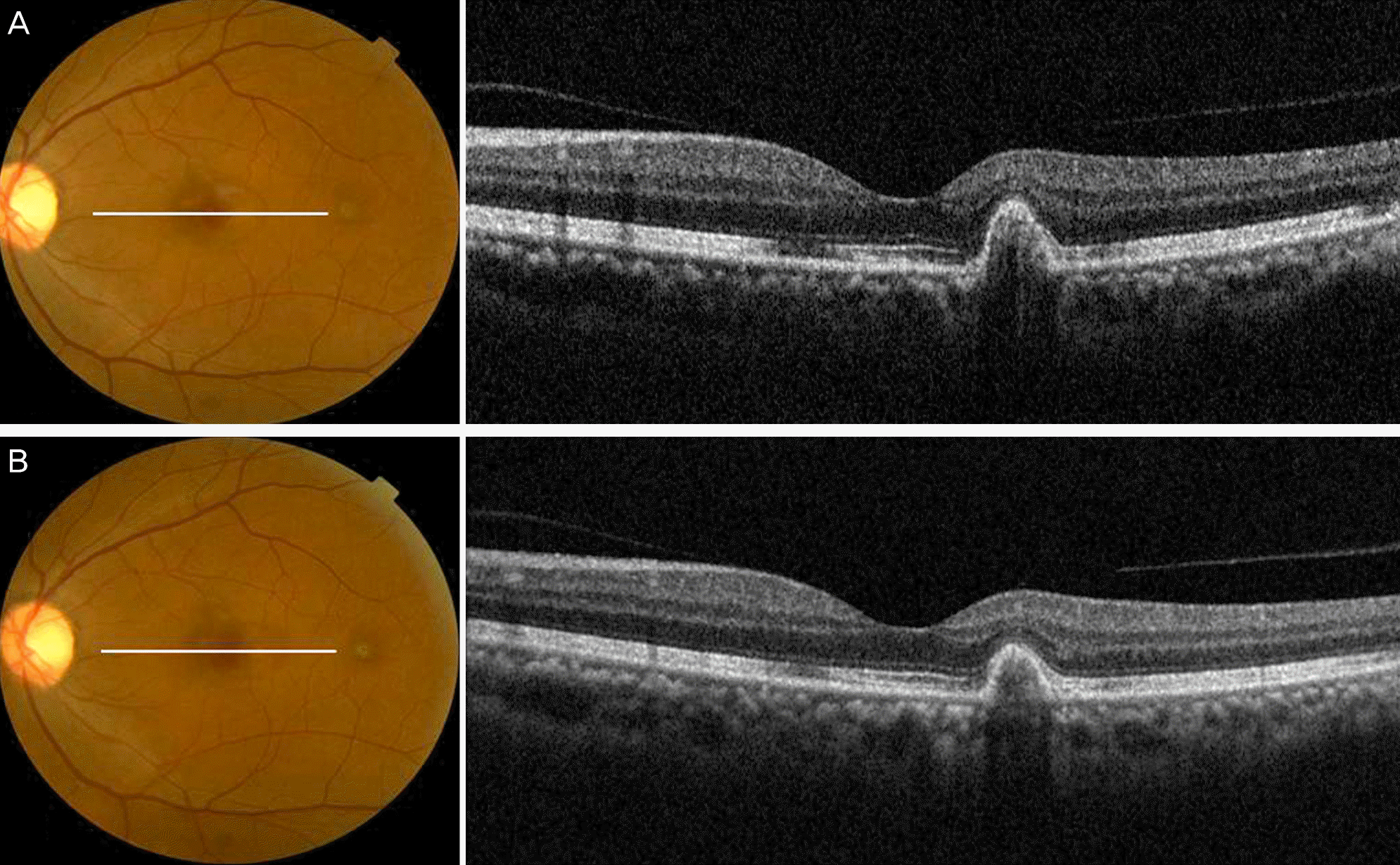
Figure 2.
(A) Color fundus and spectral-domain optical coherence tomography (OCT) images of the left eye of a patient treated with a single bevacizumab injection. Subretinal fluid and neovascular complex were not observed and only pigment epithelial detachment temporal to the fovea remained. Two additional monthly intravitreal bevacizumab injections were administered. (B) No recurrence or complications were observed on fundus photograph and OCT imaging during the eight-month follow-up.
 XML Download