Abstract
Purpose
We report a case of optic disc hemorrhage associated with buried optic nerve head drusen in a pediatric patient.
Case summary
A 10-year-old female visited our clinic with a floating sensation in her left eye, 2 days in duration. Best corrected visual acuity was 1.0 in both eyes. Intraocular pressure, light reflex, relative afferent pupillary defect and color vision were normal. The patient showed a small optic disc with blurred, irregular margins in both eyes, and optic disc hemorrhage in the left eye on fundus examination. Visual field examination revealed an enlarged blind spot in the left eye. To achieve correct diagnosis, brain MRI was performed and revealed normal findings. On spectral-domain optical coherence tomography (OCT), hyper-reflective and heterogeneous mass like lesions were found with buried optic nerve head drusen.
Go to : 
References
1. Lee KM, Woo SJ, Hwang JM. Morphologic characteristics of optic nerve head drusen on spectral-domain optical coherence tomography. Am J Ophthalmol. 2013; 155:1139–47.

2. Lee KM, Woo SJ, Hwang JM. Differentiation of optic nerve head drusen and optic disc edema with spectral-domain optical coherence tomography. Ophthalmology. 2011; 118:971–7.

3. Neffendorf JE, Mulholland C, Quinlan M, Lyons CJ. Disc drusen complicated by optic disc hemorrhage in childhood. Can J Ophthalmol. 2010; 45:537–8.

5. Sanders TE, Gay AJ, Newman M. Hemorrhagic complications of drusen of the optic disk. Am J Ophthalmol. 1971; 71:204–17.

6. Mullie MA, Sanders MD. Scleral canal size and optic nerve head drusen. Am J Ophthalmol. 1985; 99:356–9.

7. Harris MJ, Fine SL, Owens SL. Hemorrhagic complications of optic nerve drusen. Am J Ophthmol. 1981; 92:70–6.

8. Johnson LN, Diehl ML, Hamm CW, et al. Differentiating optic disc edema from optic nerve head drusen on optical coherence tomography. Arch Ophthalmol. 2009; 127:45–9.

9. Grippo TM, Shihadeh WA, Schargus M, et al. Optic nerve head drusen and visual field loss in normotensive and hypertensive eyes. J Glaucoma. 2008; 17:100–4.

10. Chaudhry NA, Lavaque AJ, Shah A, Liggett PE. Photodynamic therapy for choroidal neovascular memebrane secondary to optic nerve drusen. Ophthalmic Surg Lasers Imaging. 2005; 36:70–2.
11. Knape RM, Zavaleta EM, Clark CL 3rd, et al. Intravitreal bevacizumab treatment of bilateral peripapillary choroidal neo- vascularization from optic nerve head drusen. J AAPOS. 2011; 15:87–90.
Go to : 
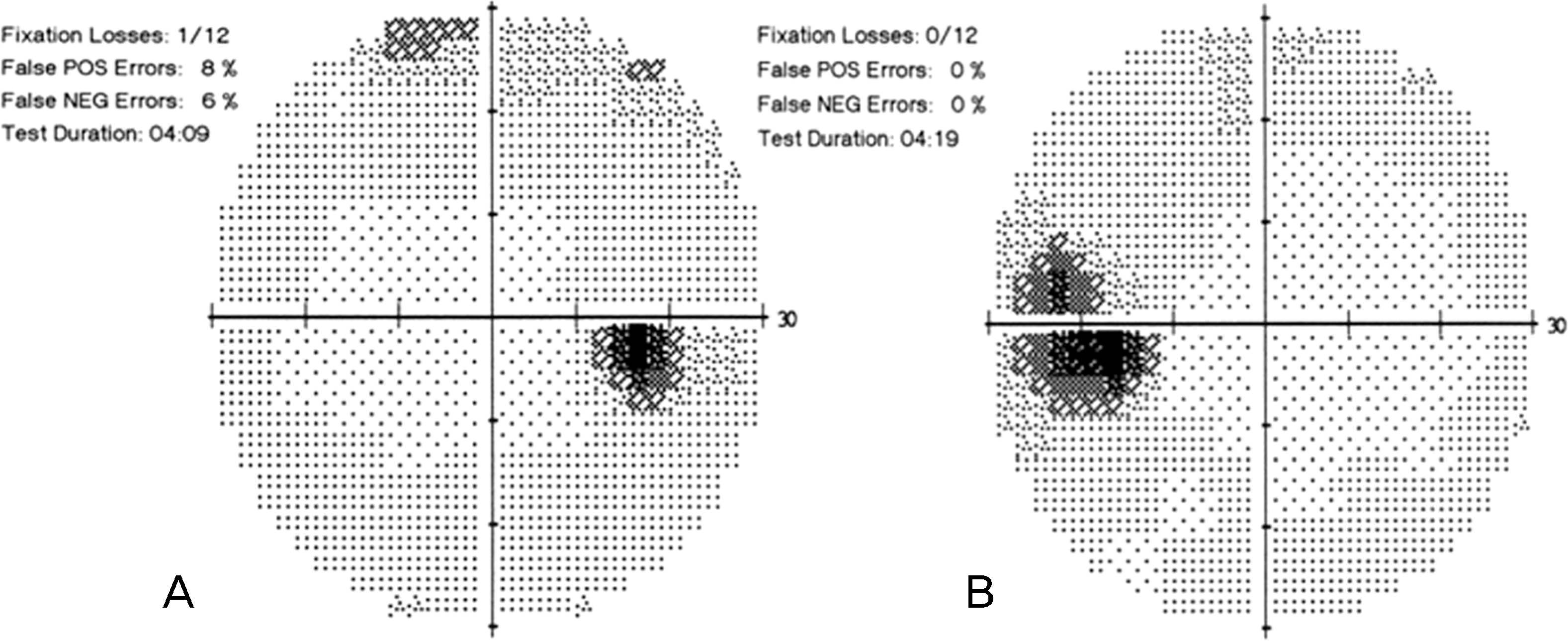 | Figure 1.Visual field tests at the first visit. These show normal in the right eye (A) and the enlargement of blind spot in the left eye (B). POS = positive; NEG = negative. |
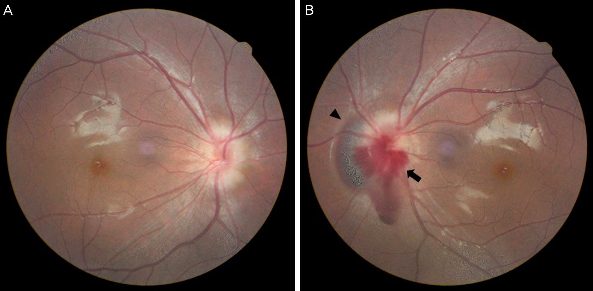 | Figure 2.Fundus photographs at the first visit (A, B). These show bilateral small optic disc with blurred, irregular margin, suggestive of optic nerve head drusen. The arrowhead indicates deep peripapillary hemorrhage. The arrow indicates splinter hemorrhage. |
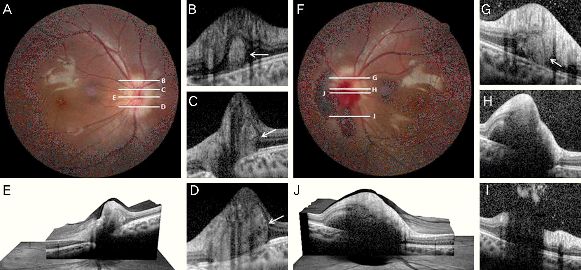 | Figure 3.Images showing multiple buried optic disc drusen within small optic disc of the right eye (A-E) and left eye (F-J). White horizontal lines in fundus photographs (A, F) indicate the horizontal cross sections level of the optical coherence tomography (OCT) scan (B-E, G-J). E and J images show three-dimensional image-OCT on center of optic disc. These reveal buried optic nerve head drusen with a subretinal hyperreflective mass like lesion (B-D, G, white arrows) with optic disc elevation in both eyes. Deep peripapillary hemorrhage and vitreous hemorrhage reflect hyporeflectivity on the subretinal and preretinal space in the left eye. |
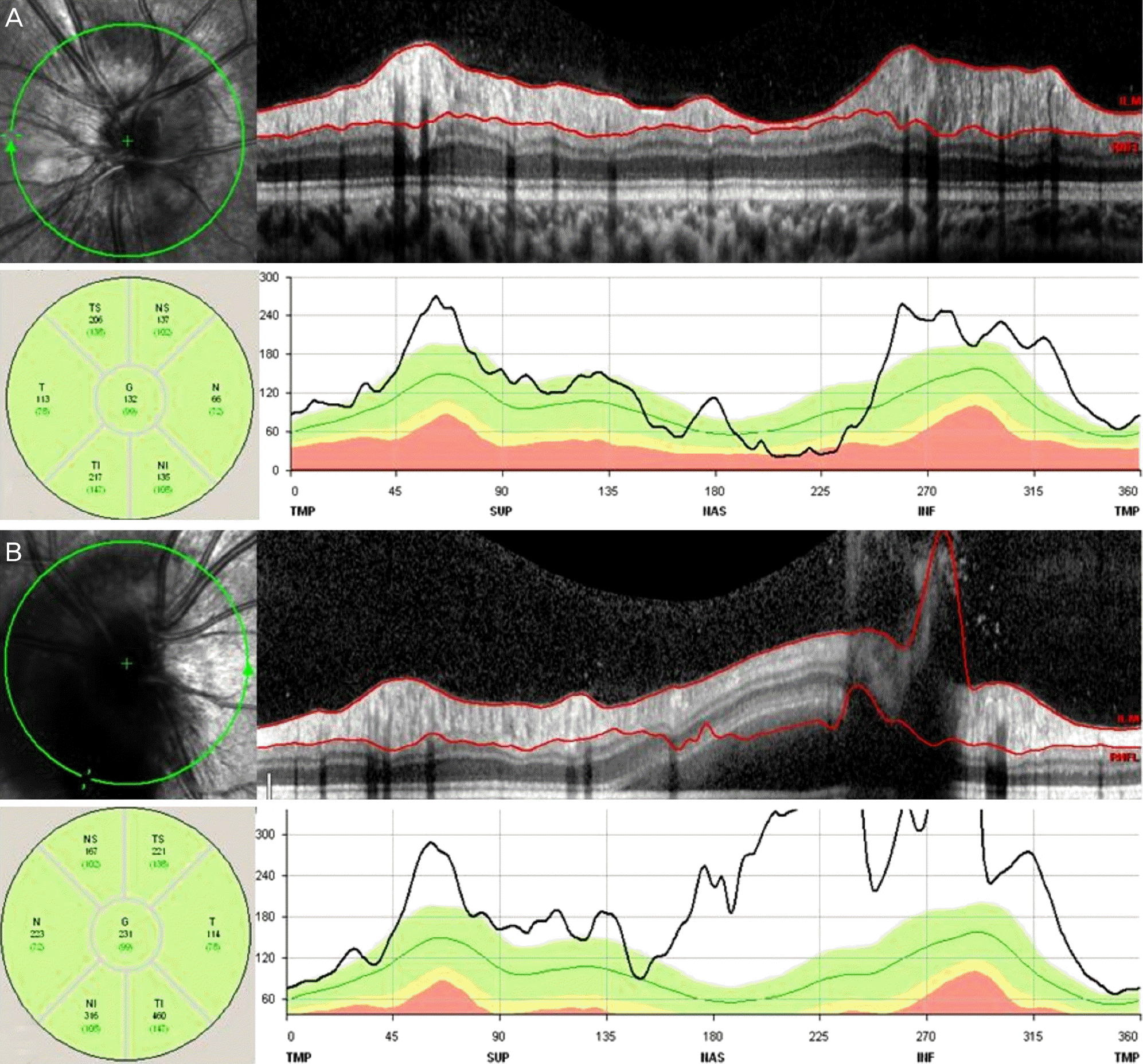 | Figure 4.Retinal nerve fiber layer (RNFL) measurement using spectralis optical coherence tomography (OCT) of both eyes. (A) The RNFL thickness is increased in all sections except for nasal section by the underlying buried optic nerve head drusen in the right eye. (B) Because of deep peripapillary hemorrhage in the left eye, the RNFL thickness cannot be measured accurately. ILM = inner limiting membrane; TS = superotemporal; NS = superonasal; T = temporal; G = general; N = nasal; TI = inferotemporal; NI = inferonasal; TMP = temporal; SUP = superior; NAS = nasal; INF = inferior. |
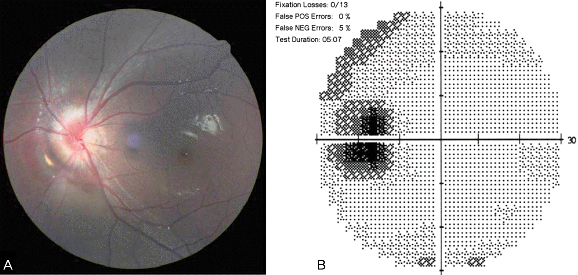 | Figure 5.Fundus photograph and visual field test after 1 month. (A) Fundus photograph shows the disappearance of peripapillary hemorrhage. (B) Visual field test shows the enlargement of blind spot, not much change comparing with visual field test of the first visit. POS = positive; NEG = negative. |
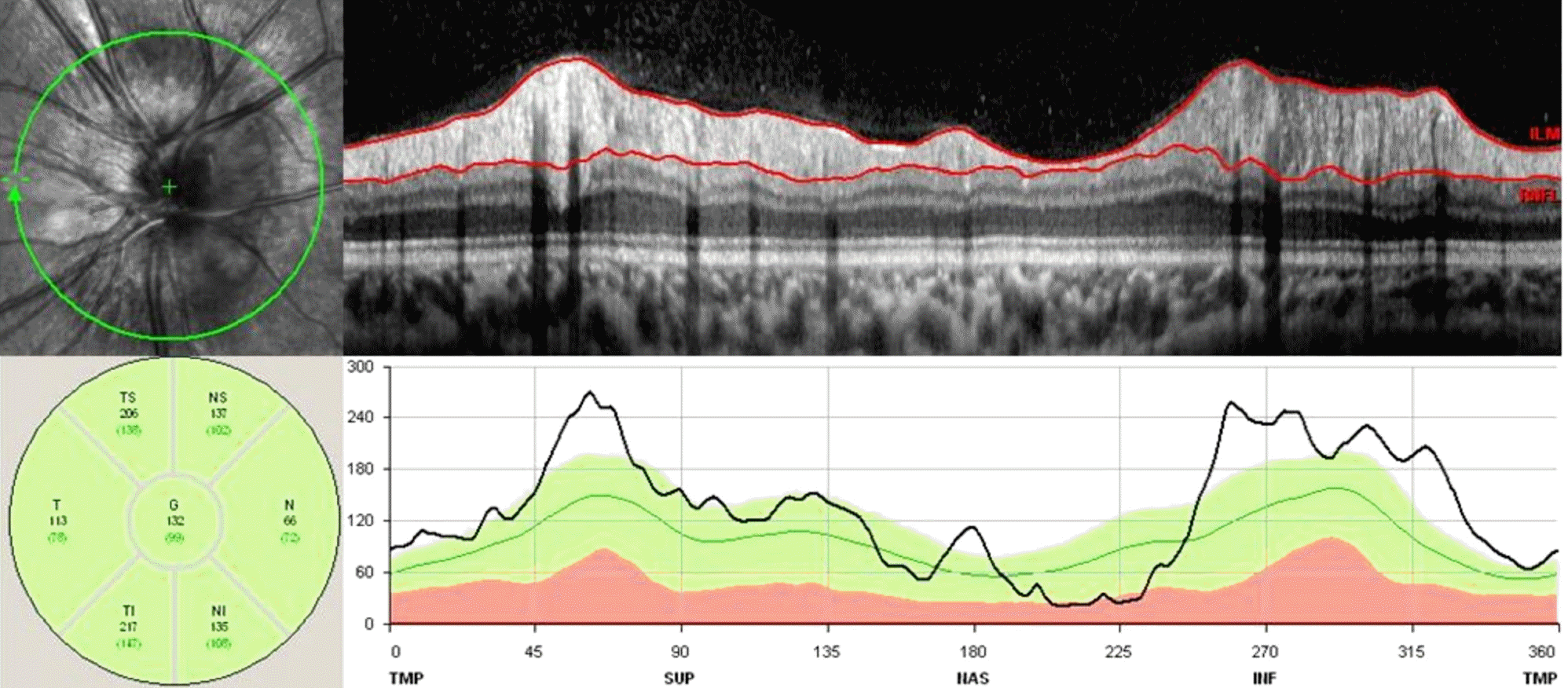 | Figure 6.Retinal nerve fiber layer (RNFL) measurement using spectralis optical coherence tomography (OCT) of the left eye after the disappearance of peripapillary hemorrhage, after 1 month. The RNFL thickness is increased in all sections except for nasal section by the underlying buried optic nerve head drusen. ILM = inner limiting membrane; TS = superotemporal; NS = superonasal; T = temporal; G = general; N = nasal; TI = inferotemporal; NI = inferonasal; TMP = temporal; SUP = superior; NAS = nasal; INF = inferior. |


 PDF
PDF ePub
ePub Citation
 Print
Print


 XML Download