

 PDF
PDF ePub
ePub Citation
 Print
Print


Abstract
Purpose
To report a patient diagnosed with adult-onset vitelliform dystrophy (AOVD) who received an intravitreal injection of bevacizumab in both eyes.
Case summary
A 47-year-old female presented with blurred vision and metamorphopsia in both eyes. On color fundus photograph, small, round, yellowish dots on the foveola and subreitnal fluid were observed. Optical coherence tomography (OCT) showed thick hyperreflective structures in the retinal pigment epithelium (RPE) layer with serous retinal detachment and subretinal fluid. Despite an intravitreal injection of bevacizumab on both eyes, anatomical improvement was not observed on fundus photography and OCT.
References
1. Do P, Ferrucci S. Adult-onset foveomacular vitelliform dystrophy. Optometry. 2006; 77:156–66.

2. Funk M, Kriechbaum K, Prager F, et al. Intraocular concentrations of growth factors and cytokines in retinal vein occlusion and the effect of therapy with bevacizumab. Invest Ophthalmol Vis Sci. 2009; 50:1025–32.
3. Wu L, Martínez-Castellanos MA, Quiroz-Mercado H, et al. Twelvemonth safety of intravitreal injections of bevacizumab (Avastin): results of the Pan-American Collaborative Retina Study Group (PACORES). Graefes Arch Clin Exp Ophthalmol. 2008; 246:81–7.
4. Kreutzer TC, Alge CS, Wolf AH, et al. Intravitreal bevacizumab for the treatment of macular oedema secondary to branch retinal vein occlusion. Br J Ophthalmol. 2008; 92:351–5.
5. Rabena MD, Pieramici DJ, Castellarin AA, et al. Intravitreal bevacizumab (Avastin) in the treatment of macular edema secondary to branch retinal vein occlusion. Retina. 2007; 27:419–25.
6. Matsumoto Y, Freund KB, Peiretti E, et al. Rebound macular edema following bevacizumab (Avastin) therapy for retinal venous occlusive disease. Retina. 2007; 27:426–31.
7. Lee JY, Lim J, Chung H, et al. Spectral domain optical coherence tomography in a patient with adult-onset vitelliform dystrophy treated with intravitreal bevacizumab. Ophthalmic Surg Lasers Imaging. 2009; 40:319–21.
8. Petrukhin K, Koisti MJ, Bakall B, et al. Identification of the gene responsible for Best macular dystrophy. Nat Genet. 1998; 19:241–7.
9. Marmorstein AD, Marmorstein LY, Rayborn M, et al. Bestrophin, the product of the Best vitelliform macular dystrophy gene (VMD2), localizes to the basolateral plasma membrane of the retinal pigment epithelium. Proc Natl Acad Sci U S A. 2000; 97:12758–63.
10. Sun H, Tsunenari T, Yau KW, Nathans J. The vitelliform macular dystrophy protein defines a new family of chloride channels. Proc Natl Acad Sci U S A. 2002; 99:4008–13.
11. Boon CJ, Klevering BJ, Leroy BP, et al. The spectrum of ocular phenotypes caused by mutation in the BEST 1 gene. Prog Retin Eye Res. 2009; 28:187–205.
12. Booij JC, Boon CJ, van SChooneveld MJ, et al. Course of visual decline in relation to the BEST1 genotype in vitelliform macular dystrophy. Ophthalmology. 2010; 117:1415–22.
13. Jarc-Vidmar M, Kraut A, Hawlina M. Fundus autofluorescence imaging in Best's vitelliform dystrophy. Klin Monatsbl Augenheilkd. 2003; 220:861–7.
14. Gass JDM. Stereoscopic atlas of macular diseases: diagnosis and treatment. 4th ed.St. Louis: Mosby;1997. p. 304–11.
15. Deutman AF, Hoyng CB. Macular dystrophies. In : Ryan SJ, Ogden TE, Hinton DR, editors. Retina. 3rd.2. St. Louis: Mosby;2001. p. 1210–57.
16. Spaide RF, Noble K, Morgan A, Freund KB. Vitelliform macular dystrophy. Ophthalmology. 2006; 113:1392–400.
17. Montero JA, Ruiz-Moreno JM, De La Vega C. Intravitreal bevacizumab for adult-onset vitelliform dystrophy: a case report. Eur J Ophthalmol. 2007; 17:983–6.
18. Abengoechea-Hernandez S, Elizalde-Montagut J, Fideliz de la Paz-Dalisay M. Photodynamic therapy in adult-onset foveomacular vitelliform dystrophy. Arch Soc Esp Oftalmol. 2007; 82:117–20.
Figure 1.
Color fundus photograph of a 47-year-old female showing a small, round, yellowish dot at the site of the foveola or a tiny honeycomb structure centrally (A, B). In fluorescence angiography reveals early blockage and late intense hyperfluorescent circles with hypofluorescent central zones (C, D).
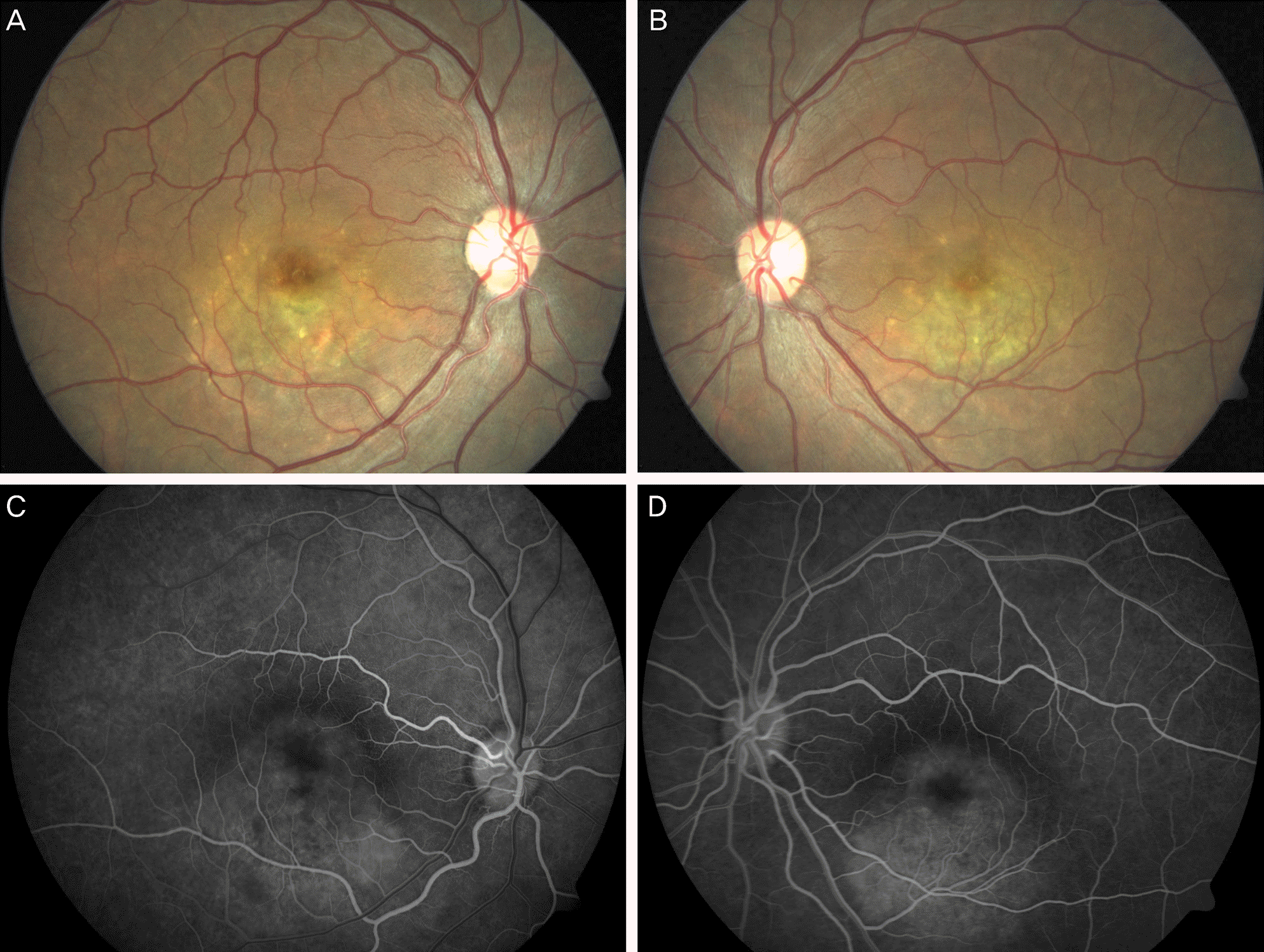
Figure 2.
Right eye optical coherence tomography (OCT) represents thick hyperreflective structures in the retinal pigment epithelium (RPE) layer and serous retinal detachment and subretinal fluid. OCT after the intravitreal bevacizumab showing no difference of subretinal fluid volume or extent of serous retinal detachment and hyperreflective lesions. IVAI = intravitreal avastin injection.

Figure 3.
Left eye optical coherence tomography (OCT) also represents abnormal findings as described above on the right eye. OCT was performed after the intravitreal bevacizumab, there were also no difference of subretinal fluid volume or extent of serous retinal detachment and hyperreflective lesions. IVAI = intravitreal avastin injection.

 XML Download