

 PDF
PDF ePub
ePub Citation
 Print
Print


Abstract
Purpose
To compare the clinical efficacy of simultaneous intracameral and intravitreal injection and intravitreal single injection of bevacizumab in patients with neovascular glaucoma (NVG).
Methods
The medical records of 43 eyes of 43 patients, who had treated with simultaneous intracameral and intravitreal injection (Group I) or intravitreal single injection (Group II) of bevacizumab 1.25 mg from January 2010 to December 2012, were retro- spectively reviewed. The best corrected visual acuity (BCVA), intraocular pressure (IOP), regression time of new vessel in the iris (NVI) and anterior chamber angle (NVA), progression of peripheral anterior synechiae (PAS), and corneal parameters were measured preoperatively and one day, three days, 1 week, 1 month, and 3 months postoperatively.
Results
There was significant changes of IOP between the two groups at 1, 3 days postoperatively (p = 0.001, p < 0.001). The regression time of NVI and NVA in Group I was significantly faster than Group II (p = 0.026, p = 0.033). In the phakic eyes, regression time of NVI and NVA was significantly longer than aphakic and pseudophakic eyes in group II (p = 0.006, p = 0.005). Also, in the phakic NVG patients, the formation of PAS in Group I was significantly less than in Group II for the postoperative three months (p = 0.020).
Conclusions
Simultaneous intravitreal and intracameral injection of bevacizumab seem to be more effective for the early lowering of IOP and regression of NVI and NVA, and inhibiting further PAS formation in NVG patients, especially in the phakic eyes. Therefore, simultaneous intracameral and intravitreal injection of bevacizumab may be considered as an adjunct to management of NVG in the phakic eyes.
Go to : 
References
1. Sivak-Callcott JA, O'Day DM, Gass JD, Tsai JC. Evidence-based recommendations for the diagnosis and treatment of neovascular glaucoma. Ophthalmology. 2001; 108:1767–76.
2. Shazly TA, Latina MA. Neovascular glaucoma: etiology, diagnosis and prognosis. Semin Ophthalmol. 2009; 24:113–21.

3. Aiello LP. Vascular endothelial growth factor and the eye: biochemical mechanisms of action and implications for novel therapies. Ophthal Res. 1997; 29:354–62.
4. Aiello LP, Avery RL, Arrigg PG, et al. Vascular endothelial growth factor in ocular fluid of patients with diabetic retinopathy and other retinal disorders. N Engl J Med. 1994; 331:1480–7.
5. Tripathi RC, Li J, Tripathi BJ, et al. Increased level of vascular endothelial growth factor in aqueous humor of patients with neovascular glaucoma. Ophthalmology. 1998; 105:232–7.
6. SooHoo JR, Seibold LK, Kahook MY. Recent advances in the management of neovascular glaucoma. Semin Ophthalmol. 2013; 28:165–72.
7. Michels S, Rosenfeld PJ, Puliafito CA, et al. Systemic bevacizumab (Avastin) therapy for neovascular age-related macular degeneration twelve-week results of an uncontrolled open-label clinical study. Ophthalmology. 2005; 112:1035–47.
8. Spaide RF, Laud K, Fine HF, et al. Intravitreal bevacizumab treatment of choroidal neovascularization secondary to age-related macular degeneration. Retina. 2006; 26:383–90.
9. Rosenfeld PJ, Fung AE, Puliafito CA. Optical coherence tomography findings after an intravitreal injection of bevacizumab (avastin) for macular edema from central retinal vein occlusion. Ophthalmic Surg Lasers Imaging. 2005; 36:336–9.
10. Iturralde D, Spaide RF, Meyerle CB. Intravitreal bevacizumab (Avastin) treatment of macular edema in central retinal vein occlusion: a short-term study. Retina. 2006; 26:279–84.
11. Spaide RF, Fisher YL. Intravitreal bevacizumab (Avastin) treatment of proliferative diabetic retinopathy complicated by vitreous hemorrhage. Retina. 2006; 26:275–8.
12. Davidorf FH, Mouser JG, Derick RJ. Rapid improvement of rubeosis iridis from a single bevacizumab (Avastin) injection. Retina. 2006; 26:354–6.
13. Silva Paula J, Jorge R, Alves Costa R, et al. Short-term results of intravitreal bevacizumab (Avastin) on anterior segment neovascularization in neovascular glaucoma. Acta Ophthalmol Scand. 2006; 84:556–7.
14. Grisanti S, Biester S, Peters S, et al. Intracameral bevacizumab for iris rubeosis. Am J Ophthalmol. 2006; 142:158–60.
15. Mason JO 3rd, Albert MA Jr, Mays A, Vail R. Regression of neovascular iris vessels by intravitreal injection of bevacizumab. Retina. 2006; 26:839–41.
16. Ilieve ME, Domig D, Wolf-Schnurrbursch U, et al. Intravitreal bevacizumab (Avastin) in the treatment of neovascular glaucoma. Am J Ophthalmol. 2006; 142:1054–6.
17. Andrijevic’-Derk B, Vatavuk Z, Bencic’ G, et al. Intravitreal bevacizumab for neovascular glaucoma. Acta Clin Croat. 2008; 47:175–9.
18. Martinez-Carpio PA, Bonafonte-Marquez E, Heredia-Garcia CD, Bonafonte-Royo S. Efficacy and safety of intravitreal injection of bevacizumab in the treatment of neovascular glaucoma: systematic review. Arch Soc Esp Oftalmol. 2008; 83:579–88.
19. L'Esperance FA. Ophthalmic lasers. 3rd ed.1. St Louis: CV Mosby Co.;1989. p. 78–112.
20. Saito Y, Higashide T, Takeda H, et al. Clinical factors related to recurrence of anterior segment neovascularization after treatment including intravitreal bevacizumab. Am J Ophthalmol. 2010; 149:964–72.
21. Brouzas D, Charakidas A, Moschos M, et al. Bevacizumab (Avastin) for the management of anterior chamber neovascularization and neovascular glaucoma. Clin Ophthalmol. 2009; 3:685–8.
22. Diabetic Retinopathy Study. Photocoagulation treatment of proliferative diabetic retinopathy: the second report of diabetic retinopathy study findings. Ophthalmology. 1978; 85:82–106.
23. Manaviat MR, Rashidi M, Afkhami-Ardekani M, et al. Effect of pan retinal photocoagulation on the serum levels of vascular endothelial growth factor in diabetic patients. Int Ophthalmol. 2011; 31:271–5.
24. Wolf A, von Jagow B, Ulbig M, Haritoglou C. Intracameral Injection of bevacizumab for the treatment of neovascular glaucoma. Ophthalmologica. 2011; 226:51–6.
25. Yuzbasioglu E, Artunay O, Rasier R, et al. Simultaneous intravitreal and intracameral injection of bevacizumab (Avastin) in neovascular glaucoma. J Ocul Pharmacol Ther. 2009; 25:259–64.
26. Ghanem AA, El-Kannishy AM, El-Wehidy AS, El-Agamy AF. Intravitreal bevacizumab (avastin) as an adjuvant treatment in cases of neovascular glaucoma. Middle East Afr J Ophthalmol. 2009; 16:75–9.
27. Bakri SJ, Snyder MR, Reid JM, et al. Pharmacokinetics of intra- vitreal bevacizumab (Avastin). Ophthalmology. 2007; 114:855–9.
28. Matsuyama K, Oqata N, Jo N, et al. Levels of vascular endothelial growth factor and pigment epithelium-derived factor in eyes before and after intravitreal injection of bevacizumab. Jpn J Ophthalmol. 2009; 53:243–8.
29. Sheng YH. Vitreous prolapse during cataract surgery. Zhonghua Yan Ke Za Zhi. 1993; 29:27–9.
30. Larsson L, Osterlin S. Retinal vessels in the ora region. Possible role in the vitreo-retinal pathology in aphakia. Acta Ophthalmology. 1981; 59:526–31.
31. Sato T, Morita S, Bando H, et al. Early vitreous hemorrhage after vitrectomy with preoperative intravitreal bevacizumab for pro- liferative diabetic retinopathy. Middle East Afr J Ophthalmol. 2013; 20:51–5.
32. Kuiper EJ, Van Nieuwenhoven FA, de Smet MD, et al. The angio-fibrotic switch of VEGF and CTGF in proliferative diabetic retinopathy. PLoS One. 2008; 3:e2675.
33. Widder RA, Lemmen KD, Dietlein TS. Neovascular glaucoma. Klin Monbl Augenheikd. 2010; 227:R15–27.
34. Ehlers JP, Spirn MJ, Lam A, et al. Combination intravitreal bevacizumab/panretinal photocoagulation versus panretinal photo- coagulation alone in the treatment of neovascular glaucoma. Retina. 2008; 28:696–702.
35. Rusovici R, Sakhalkar M, Chalam KV. Evaluation of cytotoxicity of bevacizumab on VEGF-enriched corneal endothelial cells. Mol Vis. 2011; 17:3339–46.
36. Chalam KV, Agarwal S, Brar VS, et al. Evaluation of cytotoxic effects of bevacizumab on human corneal cells. Cornea. 2009; 28:328–33.
37. Suh SY, Lee JH, Jun RM. Corneal endothelial change after intravitreal bevacizumab injection. J Korean Ophthalmol Soc. 2010; 51:1549–53.
Go to :
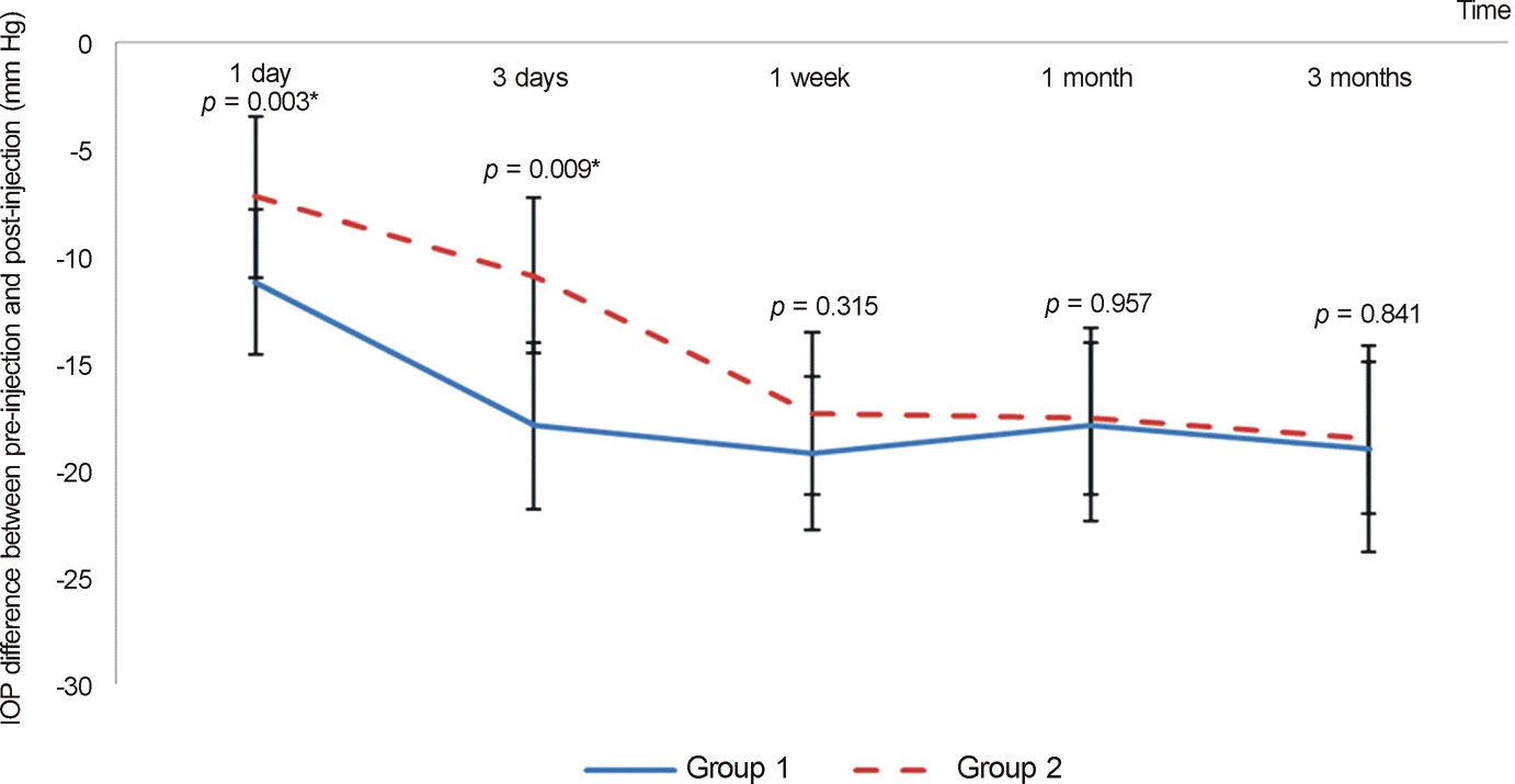 | Figure 1.IOP difference between post-injection and pre-injection in both groups. Group I: simultaneous intravitreal and intracameral bevacizumab injection group; Group II: intravitreal bevacizumab injection group. IOP = intraocular pressure. *Paired t-test, p-value < 0.05. |
Table 1.
Demographics of enrolled patients
| Group I (n = 23) | Group II (n = 21) | p-value | Total (n = 44) | |
|---|---|---|---|---|
| Age (years) | 58.7 ± 9.9 | 60.5 ± 8.8 | 0.556* | 59.9 ± 9.2 |
| Sex (n) | ||||
| Male | 14 | 16 | 30 | |
| Female | 9 | 5 | 0.731† | 14 |
| Underlying diseases (n) | ||||
| DM | 14 | 14 | 28 | |
| HTN | 3 | 2 | 5 | |
| DM and HTN | 6 | 5 | 0.668† | 11 |
| Preoperative diagnosis (n) | ||||
| PDR | 16 | 17 | 33 | |
| CRVO | 7 | 4 | 0.672† | 11 |
| Lens status (n) | ||||
| Phakia | 12 | 12 | 24 | |
| Pseudophakia | 9 | 8 | 17 | |
| Aphakia | 2 | 1 | 0.805† | 3 |
| PAS (n) | ||||
| None | 11 | 8 | 20 | |
| Mild | 4 | 1 | 5 | |
| Moderate | 3 | 1 | 4 | |
| Severe | 2 | 5 | 7 | |
| Very severe | 3 | 5 | 0.374† | 8 |
| Ahmed valve implant surgery (n) | 5 | 7 | 0.425* | 12 |
Values are presented as mean ± SD; Group I: simultaneous intravitreal and intracameral bevacizumab injection group; Group II: intravitreal bevacizumab injection group; PAS range: none = 0°, 0° < mild ≦ 90°, 90° < moderate ≦ 180°, 180° < severe ≤ 270°, 270° < very severe ≦ 360°.
Table 2.
Change of visual acuity between pre-injection and post-injection 3 months later in both groups
| Group I (n = 23) | Group II (n = 21) | p-value† | Total (n = 44) | |
|---|---|---|---|---|
| Better (n) | 4 | 5 | 9 | |
| Same (n) | 18 | 15 | 33 | |
| Worse (n) | 1 | 1 | 2 | |
| p-value* | 0.658 | 0.624 | 0.638 | |
| Δ VA (mean ± SD, n) | 0.22 ± 0.36 | 0.24 ± 0.32 | 0.882 | 0.23 ± 0.34 |
Group I: simultaneous intravitreal and intracameral bevacizumab injection group; Group II: intravitreal bevacizumab injection group.
Table 3.
Change of IOP and number of IOP lowering medications in pre-injection and 3 months after injection in both groups
| Group I(*n = 18) | Group II(*n = 14) | p-value | Total (n = 32) | ||
|---|---|---|---|---|---|
| IOP (mm Hg) | Pre-op | 36.0 ± 4.1 | 34.9 ± 4.2 | 0.637 | 35.4 ± 7.6 |
| Post-op 1 day | 26.5 ± 4.8 | 29.3 ± 3.1 | 0.385 | 27.9 ± 7.6 | |
| Post-op 3 days | 20.1 ± 3.4 | 27.4 ± 3.3 | 0.042† | 23.7 ± 7.8 | |
| Post-op 1 week | 18.8 ± 3.7 | 23.0 ± 3.2 | 0.208 | 20.9 ± 7.7 | |
| Post-op 1 month | 15.9 ± 3.6 | 16.0 ± 3.8 | 0.935 | 16.0 ± 3.6 | |
| Post-op 3 months | 14.4 ± 2.5 | 16.0 ± 2.4 | 0.230 | 15.2 ± 2.6 | |
| IOP lowering medications (n) | Pre-op | 3.00 ± 0.0 | 2.91 ± 0.3 | 0.164 | 2.96 ± 0.2 |
| Post-op 3 months | 2.47 ± 0.4 | 2.36 ± 1.1 | 0.189 | 2.42 ± 0.6 |
Table 4.
Comparison of regression time of NVI and NVA in both groups
| Group I (n = 23) | Group II (n = 21) | p-value | Total (n = 44) | |
|---|---|---|---|---|
| Regression time of NVI (days) | 3.1 ± 1.2 | 3.9 ± 1.6 | 0.026* | 3.5 ± 1.5 |
| Regression time of NVA (days) | 3.2 ± 1.4 | 4.0 ± 1.5 | 0.033* | 3.6 ± 1.5 |
Table 5.
Comparison of regression time of NVI and NVA according to lens status in both groups
| Phakia (Group I: n = 12, Group II: n = 12) | Pseudophakia (Group I: n = 9, Group II: n = 8) | Aphakia (Group I: n = 2, Group II: n = 1) | p-value‡ | ||
|---|---|---|---|---|---|
| Regression time of NVI (day) | Group I | 3.2 ± 1.4 | 3.1 ±1.0 | 2.0 ± 0.5 | 0.291 |
| Group II | 4.2 ± 1.9 | 3.3 ± 1.3 | 2.0 ± 0.0 | 0.006† | |
| p-value* | 0.01† | 0.565 | 1.000 | ||
| Regression time of NVA (day) | Group I | 3.2 ± 1.3 | 3.1 ± 1.4 | 2.0 ± 0.5 | 0.242 |
| Group II | 4.2 ± 1.9 | 3.4 ± 1.5 | 2.0 ± 0.0 | 0.005† | |
| p-value* | 0.009† | 0.549 | 1.000 |
Table 6.
Change of corneal parameters in pre-injection and 3 months after injection in both groups
| Group I (*n = 18) | Group II (*n = 14) | p-value† | Total (n = 32) | ||
|---|---|---|---|---|---|
| ECC | Pre | 2380.60 ± 278.95 | 2444.8 ± 360.50 | 0.342 | 2389.20 ± 322.42 |
| Post | 2264.65 ± 257.45 | 2377.05 ± 360.95 | 0.442 | 2319.85 ± 310.20 | |
| p-value† | 0.228 | 0.364 | 0.569 | ||
| Δ ECC | 77.95 | 67.75 | 0.064 | ||
| CV | Pre | 17.50 ± 5.87 | 18.55 ± 3.76 | 0.537 | 18.03 ± 4.90 |
| Post | 15.60 ± 5.42 | 16.75 ± 5.76 | 0.418 | 16.18 ± 5.70 | |
| p-value† | 0.210 | 0.256 | 0.309 | ||
| Δ CV | 1.9 | 1.8 | 0.602 | ||
| Hexa | Pre | 51.89 ± 4.19 | 53.39 ± 2.65 | 0.290 | 52.64 ± 3.54 |
| Post | 51.20 ± 4.18 | 52.64 ± 3.60 | 0.349 | 51.91 ± 3.92 | |
| p-value† | 0.685 | 0.630 | 0.627 | ||
| Δ Hexa | 0.69 | 0.75 | 0.598 | ||
| Pachy | Pre | 620.64 ± 38.53 | 625.18 ± 26.48 | 0.671 | 622.80 ± 32.85 |
| Post | 566.55 ± 36.75 | 573.81 ± 26.65 | 0.615 | 569.03 ± 31.80 | |
| p-value† | 0.014 | 0.036 | 0.015 | ||
| Δ Pachy | 54.09 | 51.37 | 0.762 |
Values are presented as mean ± SD; Group I: simultaneous intravitreal and intracameral bevacizumab injection group; Group II: intravitreal bevacizumab injection group.
Table 7.
PAS change according to lens status in both groups
| Phakia(Group I: n = 12 Group II: n = 12) | Pseudophakia(Group I: n = 9 Group II: n = 8) | Aphakia(Group I: n = 2 Group II: n = 1) | p-value‡ | Total (n = 44) | ||
|---|---|---|---|---|---|---|
| Preop PAS | Group I | 1.12 ± 0.45 | 1.00 ± 0.56 | 3.00 ± 0.00 | 0.112 | 2.12 ± 0.74 |
| Group II | 1.22 ± 0.76 | 2.42 ± 0.66 | 4.00 ± 0.00 | 0.142 | 3.64 ± 0.82 | |
| Postop PAS | Group I | 1.83 ± 0.52 | 1.69 ± 0.58 | 3.00 ± 0.00 | 0.346 | 3.52 ± 0.61 |
| Group II | 2.03 ± 0.82 | 3.14 ± 0.64 | 4.00 ± 0.00 | 0.052 | 5.17 ± 0.73 | |
| Δ PAS | Group I | 0.71 ± 0.12 | 0.69 ± 0.09 | 0 | 0.650 | 0.70 ± 0.18 |
| Group II | 0.81 ± 0.18 | 0.72 ± 0.12 | 0 | 0.262 | 0.76 ± 0.15 | |
| p-value* | 0.020† | 0.317 | 0.068 |
Values are presented as mean ± SD; Group I: simultaneous intravitreal and intracameral bevacizumab injection group; Group II: intravitreal bevacizumab injection group.
 XML Download