

 PDF
PDF ePub
ePub Citation
 Print
Print


Abstract
Purpose
To report a case of secondary pigmentary glaucoma after implantable contact lens (ICL) implantation success-fully treated with trabeculectomy without ICL removal.
Case summary
A 29-year-old woman presented with refractory intraocular pressure (IOP) increase in both eyes. IOP was 22 mm Hg in the right eye and 39 mm Hg in the left eye. The patient received posterior chamber phakic intraocular lens im-plantation in both eyes 22 months prior. Slit lamp examination revealed patent iridotomy sites in both eyes. Gonioscopy re-vealed open angles with 4-degree pigment deposits on the trabecular meshwork in both eyes. Ultrasound biomicroscopy examination confirmed contact between ICL and the posterior surface of the iris. In spite of well tolerated medical therapy and selective laser trabeculoplasty, IOP was 46 mm Hg in her left eye. Trabeculectomy was performed in her left eye with-out ICL removal. At 6 months postoperative, IOP measured 6 mm Hg without any anti-glaucoma medication and bleb was maintained in good condition in the left eye.
Go to : 
References
1. Fyodorov SN, Zuyev VK, Aznabayev BM. Intraocular correction of high myopia with negative posterior chamber lens. Ophthalmosurg. 1991; 3:57–8.
2. Lee SY, Cheon HJ, Baek TM, Lee KH. Implantable contact lens to correct high myopia (clinical study with 24 months follow-up). J Korean Ophthalmol Soc. 2000; 41:1515–22.
3. Rosen E, Gore C. Staar Collamer posterior chamber phakic intra-ocular lens to correct myopia and hyperopia. J Cataract Refract Surg. 1998; 24:596–606.

4. Khan AJ, Percival SP. 12 year results of a prospective trial comparing poly(methyl methacrylate) and poly(hydroxyethyl methacrylate) intraocular lenses. J Cataract Refract Surg. 1999; 25:1404–7.
5. Chung YW, Byun YS, Chung SK. Long-term changes in tilt, de-centration and anterior chamber depth after implantable collamer lens insertion. J Korean Ophthalmol Soc. 2011; 52:157–62.
6. Ibrahim O, Waring GO 3rd. Successful exchange of dislocated phakic intraocular lens. J Refract Surg. 1995; 11:282–3.
7. Chun YS, Park IK, Lee HI. . Iris and trabecular meshwork pig-ment changes after posterior chamber phakic intraocular lens implantation. J Cataract Refract Surg. 2006; 32:1452–8.
8. Sánchez-Galeana CA, Zadok D, Montes M. . Refractory intra-ocular pressure increase after phakic posterior chamber intraocular lens implantation. Am J Ophthalmol. 2002; 134:121–3.
9. Brandt JD, Mockovak ME, Chayet A. Pigmentary dispersion syndrome induced by a posterior chamber phakic refractive lens. Am J Ophthalmol. 2001; 131:260–3.
10. Farrar SM, Shields MB. Current concepts in pigmentary glaucoma. Surv Ophthalmol. 1993; 37:233–52.
11. Campbell DG. Pigmentary dispersion and glaucoma: a new theory. Arch Ophthalmol. 1979; 97:1667–72.
12. Han SY, Lee KH. Long term effect of ICL implantation to treat high myopia. J Korean Ophthalmol Soc. 2007; 48:465–72.
13. Abela-Formanek C, Kruger AJ, Dejaco-Ruhswurm I. . Gon- ioscopic changes after implantation of a posterior chamber lens in phakic myopic eyes. J Cataract Refract Surg. 2001; 27:1919–25.
14. Mitchell P, Hourihan F, Sandbach J, Wang JJ. The relationship be-tween glaucoma and myopia: the Blue Mountains Eye Study. Ophthalmology. 1999; 106:2010–5.
15. Zaldivar R, Davidorf JM, Oscherow S. Posterior chamber phakic intraocular lens for myopia of -8 to -19 diopters. J Refract Surg. 1998; 14:294–305.
16. Farrar SM, Shields MB. Current concepts in pigmentary glaucoma. Surv Ophthalmol. 1993; 37:233–52.
17. Damji KF, Bovell AM, Hodge WG. . Selective laser trabeculo-plasty versus argon laser trabeculoplasty: results from a 1-year randomised clinical trial. Br J Ophthalmol. 2006; 90:1490–4.
Go to :
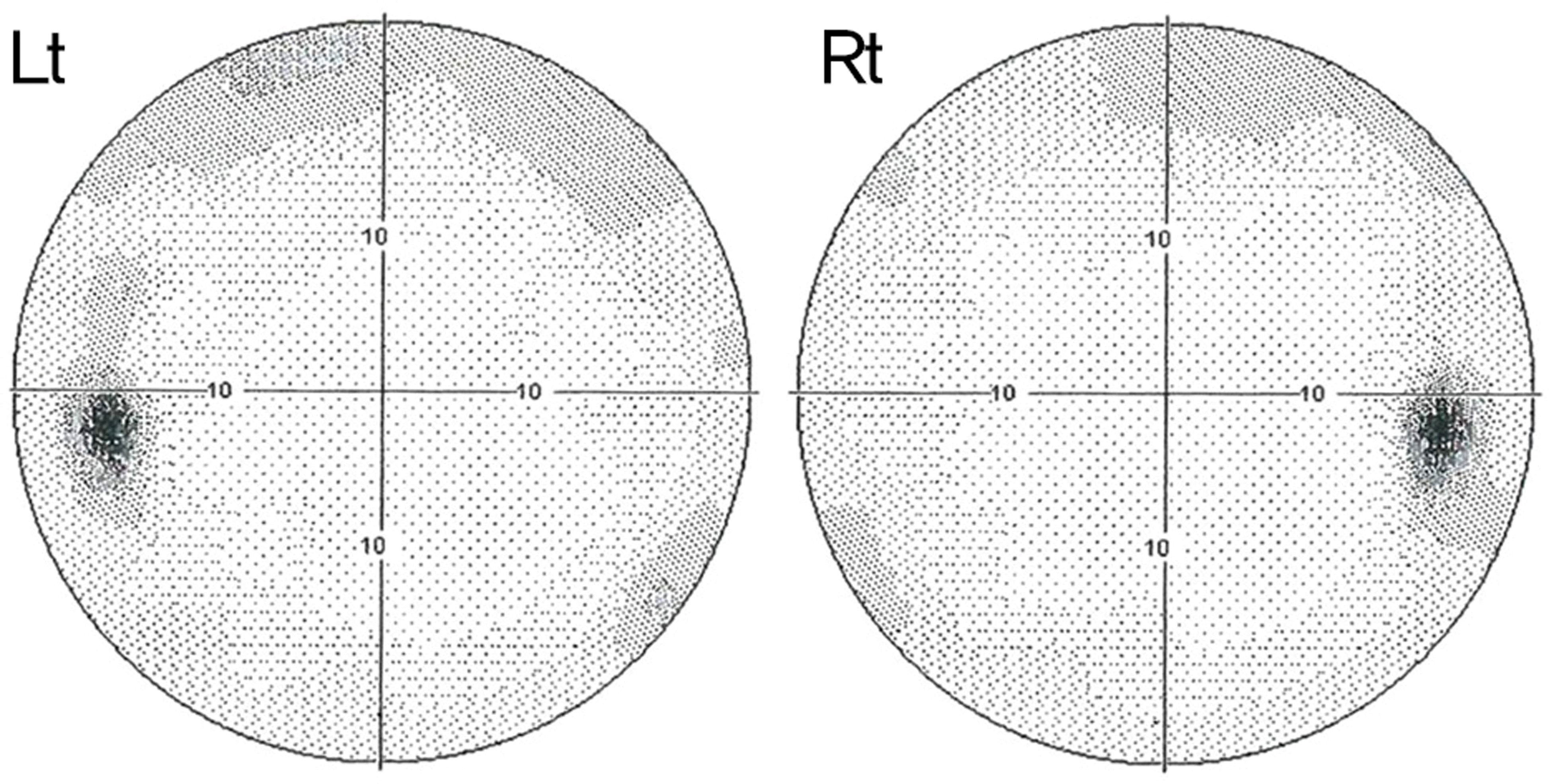 | Figure 1.Automated visual field examination before ICL im-plantation shows no glaucomatous visual field defects in both eyes. |
 | Figure 2.(A) Slit lamp photograph at the initial visit shows patent laser iridotomy sites (white block arrows) at the 10 and 2 o’clock positions. (B) Gonioscopy reveals open angles with 4 degrees of pigment deposited on the trabecular meshwork (black arrow heads) and forward vaulting of the iris overlying the ICL. (C) Ultrasound biomicoscopy examination confirms contact and rubbing between ICL and posterior surface of the iris (white open block arrows). |
 | Figure 3.(A) Disc photograph shows vertical cup-disc ratio of 0.75 in the right eye and 0.8 in the left eye consistent with glaucoma-tous optic disc change. (B) Red-free fundus photography shows superior and inferior retinal nerve fiber layer (RNFL) defects. (C) Visual field examination reveals nasal step defect in the right eye and superior arcuate with inferior nasal step defect in the left eye. (D) Optical coherence tomography shows RNFL thinning in the superotemporal and inferotemporal region in TSNIT graph con-sistent with visual field examinations. |

 XML Download