

 PDF
PDF ePub
ePub Citation
 Print
Print


Abstract
Purpose
To compare the efficacy of time domain (TD) and spectral domain (SD) optical coherence tomography (OCT) in determining vitreomacular interface (VMI).
Methods
VMIs were evaluated with TD and SD OCT images crossing the fovea horizontally in 69 eyes (mean age 52.7 ± 15.4 years) and were classified as follows: (1) no vitreomacular separation (VMS), (2) incomplete VMS, and (3) unknown.
Results
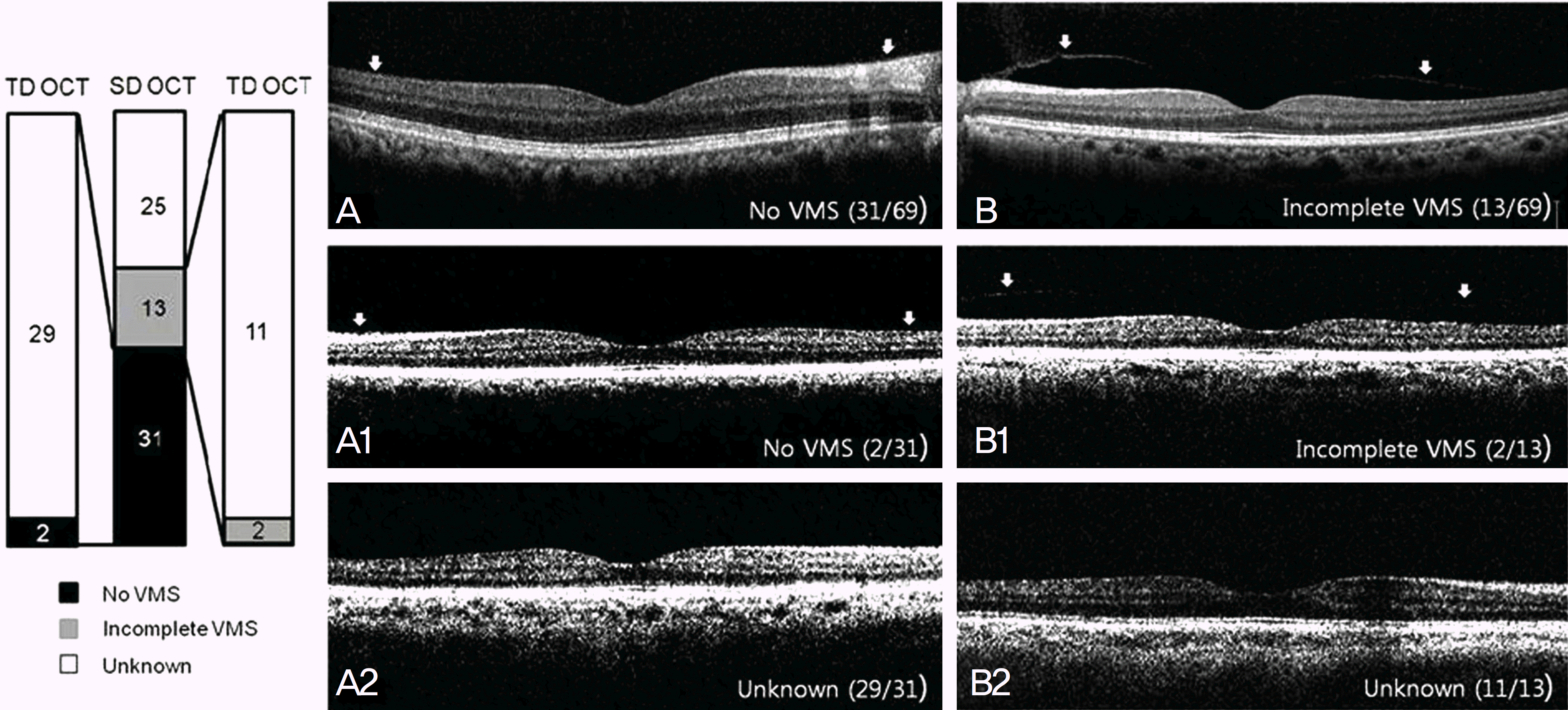
In TD OCT, no VMS was observed in 2 eyes (2.9%), incomplete VMS in 2 eyes (2.9%), and unknown in 65 eyes (94.2%). In SD OCT, no VMS was observed in 31 eyes (45.0%), incomplete VMS in 13 eyes (18.8%), and unknown in 25 eyes (36.2%). In 31 eyes with no VMS on SD OCT, 29 eyes (93.5%) presented unknown on TD OCT (p<0.0001). In 13 eyes with incomplete VMS on SD OCT, 2 eyes (15.4%) showed incomplete VMS and 11 eyes (84.6%) showed unknown on TD OCT (p<0.0001). TD OCT was also non-informative in all 25 eyes with unknown on SD OCT.
References
1. Patronas M, Kroll AJ, Lou PL, Ryan EA. A review of vitreoretinal interface pathology. Int Ophthalmol Clin. 2009; 49:133–43.

2. Voo I, Mavrofrides EC, Puliafito CA. Clinical applications of abdominal coherence tomography for the diagnosis and management of macular diseases. Ophthalmol Clin North Am. 2004; 17:21–31.
3. Fisher YL, Slakter JS, Friedman RA, Yannuzzi LA. Kinetic ultrasound evaluation of the posterior vitreoretinal interface. Ophthalmology. 1991; 98:1135–8.
4. Hoehn F, Mirshahi A, Hattenbach LO. Optical coherence abdominal for diagnosis of posterior vitreous detachment at the macular region. Eur J Ophthalmol. 2009; 19:442–7.
5. Huang D, Swanson EA, Lin CP, et al. Optical coherence tomography. Science. 1991; 254:1178–81.
6. Tammewar AM, Bartsch DU, Kozak I, et al. Imaging abdominal interface abnormalities in the coronal plane by abdominal combined scanning laser and optical coherence tomography. Br J Ophthalmol. 2009; 93:366–72.
7. Drexler W, Fujimoto JG. State-of-the-art retinal optical coherence tomography. Prog Retin Eye Res. 2008; 27:45–88.
8. Luviano DM, Benz MS, Kim RY, et al. Selected clinical abdominals of spectral domain and time domain optical coherence tomography. Ophthalmic Surg Lasers Imaging. 2009; 40:325–8.
9. Yi K, Chen TC, de Boer JF. Spectral domain optical coherence tomography. Techniques in Ophthalmology. 2006; 4:170–4.
10. Mirza RG, Johnson MW, Jampol LM. Optical coherence abdominal use in evaluation of the vitreoretinal interface: a review. Surv Ophthalmol. 2007; 52:397–421.
11. Brennen PM, Kagemann L, Friberg TR. Comparison of Stratus OCT and Cirrus HD-OCT imaging in macular diseases. Ophthalmic Surg Lasers Imaging. 2009; 40:25–31.
12. Nigam N, Bartsch DU, Cheng L, et al. Spectral domain optical abdominal tomography for imaging ERM, retinal edema, and abdominal interface. Retina. 2010; 30:246–53.
13. Ishikawa H, Gürses-Ozden R, Hoh ST, et al. Grayscale and pro-portion-corrected optical coherence tomography images. Ophthalmic Surg Lasers. 2000; 31:223–8.
14. Perichon JY, Brasseur G, Uzzan J. [Ultrasonographic study of abdominal vitreous detachment in emmetropic eyes]. J Fr Ophthalmol. 1993; 16:538–44.
15. Zeimer R, Shahidi M, Mori M, et al. A new method for rapid abdominal of the retinal thickness at the posterior pole. Invest Ophthalmol Vis Sci. 1996; 37:1994–2001.
16. Legarreta JE, Gregori G, Punjabi OS, et al. Macular thickness measurements in normal eyes using spectral domain optical abdominal tomography. Ophthalmic Surg Lasers Imaging. 2008; 39(4 Suppl):S43–9.
17. Uchino E, Uemura A, Ohba N. Initial stages of posterior vitreous detachment in healthy eyes of older persons evaluated by optical coherence tomography. Arch Ophthalmol. 2001; 119:1475–9.
18. Johnson MW. Posterior vitreous detachment: evolution and abdominals of its early stages. Am J Ophthalmol. 2010; 149:371–82. e1.
19. Gaudric A, Haouchine B, Massin P, et al. Macular hole formation: new data provided by optical coherence tomography. Arch Ophthalmol. 1999; 117:744–51.
20. Weber-Krause B, Eckardt C. [Incidence of posterior vitreous abdominal in the elderly]. Ophthalmologe. 1997; 94:619–23.
21. Yonemoto J, Ideta H, Sasaki K, et al. The age of onset of posterior vitreous detachment. Graefes Arch Clin Exp Ophthalmol. 1994; 232:67–70.
22. Kishi S, Demaria C, Shimizu K. Vitreous cortex remnants at the fovea after spontaneous vitreous detachment. Int Ophthalmol. 1986; 9:253–60.
23. Gupta P, Yee KM, Garcia P, et al. Vitreoschisis in macular diseases. Br J Ophthalmol. 2011; 95:376–80.
Figure 1.
No VMS: SD OCT image of macula showing complete attachment of the posterior hyaloid to the retinal surface (arrows).
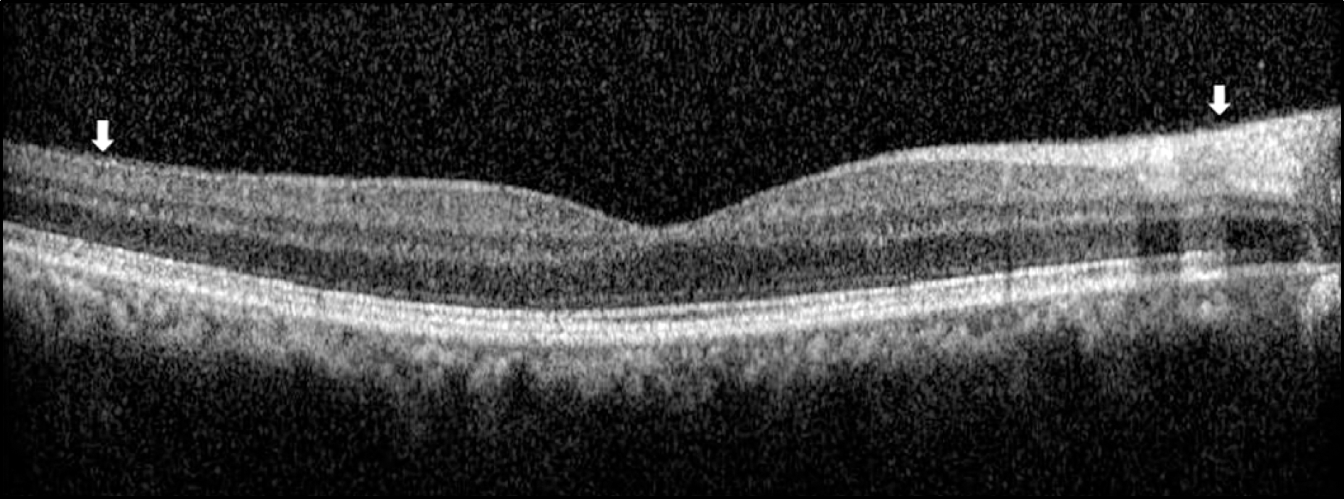
Figure 2.
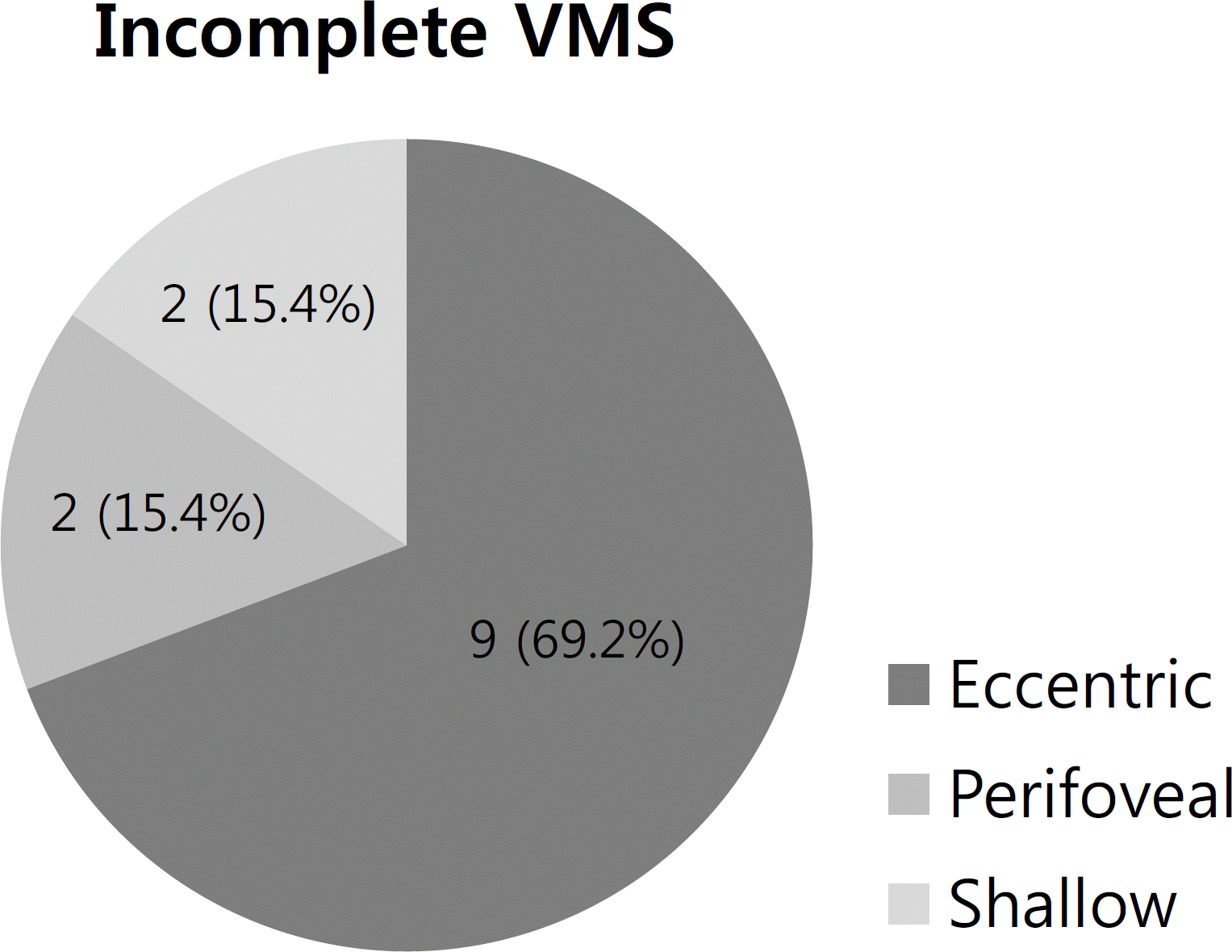
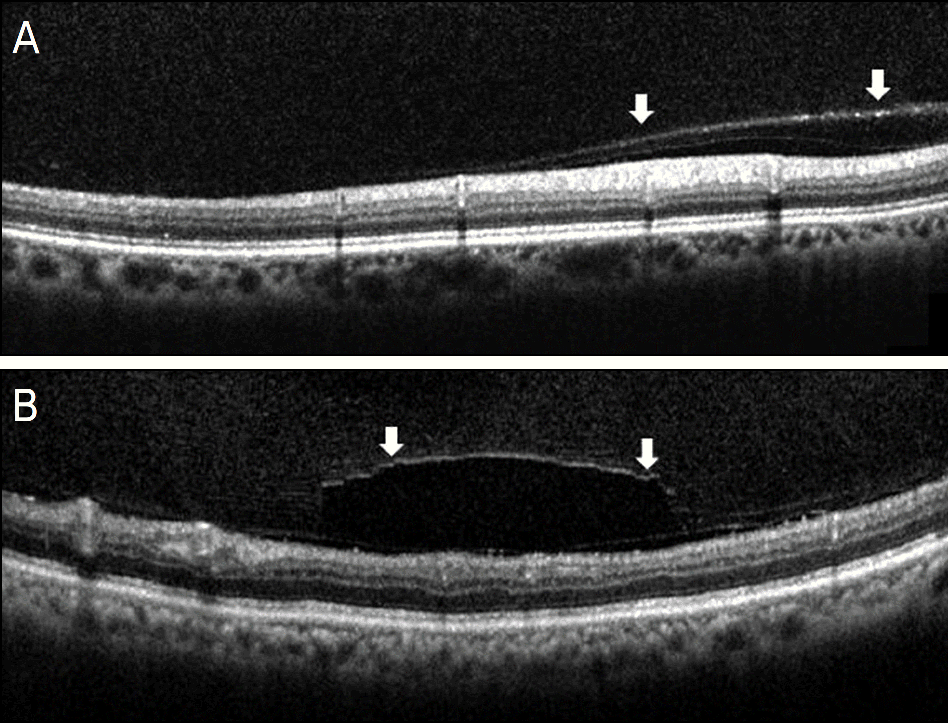
Incomplete VMS: SD OCT images for eccentric VMS (A), and perifoveal VMS (B), and shallow VMS (C).
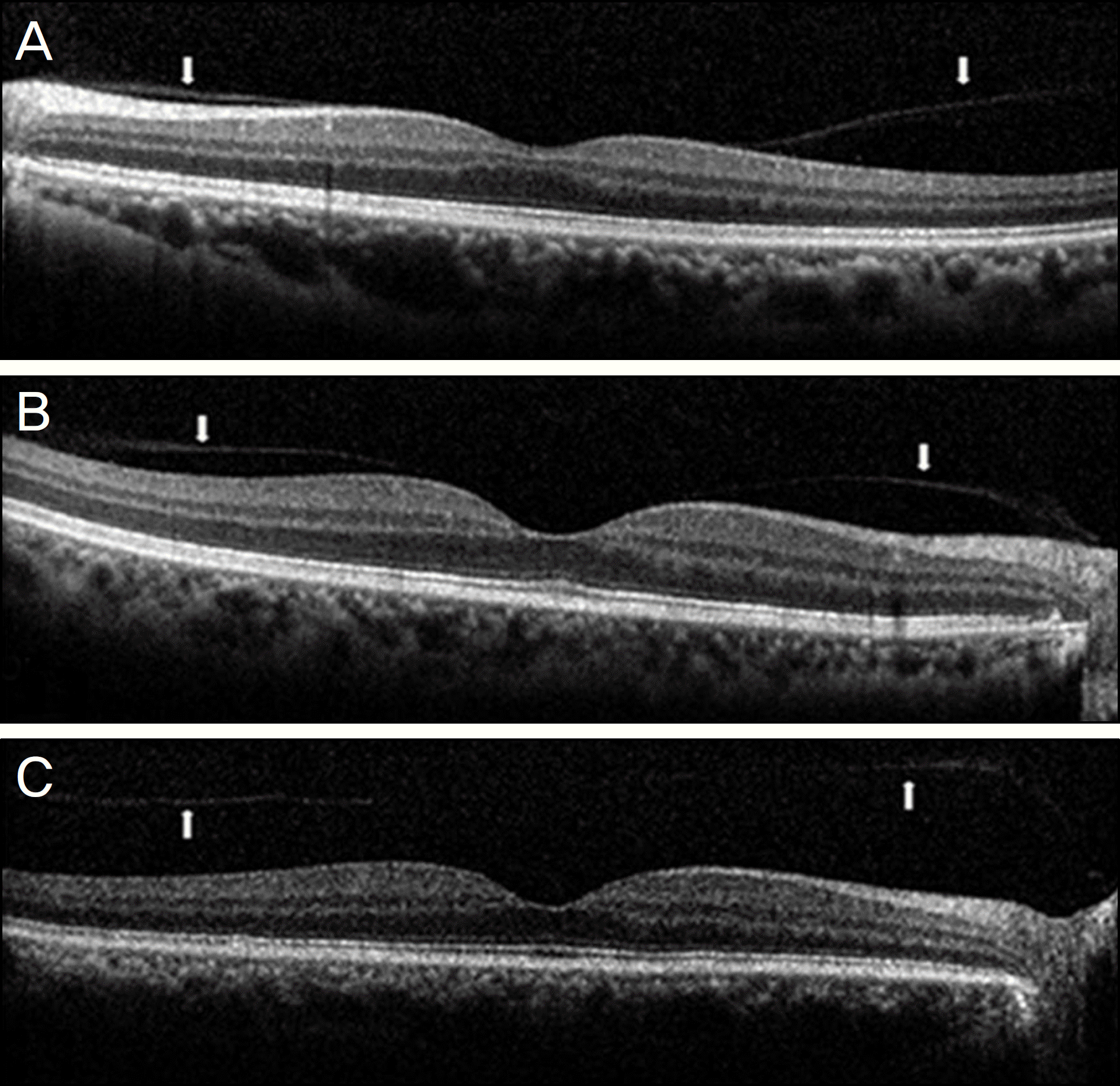
Figure 4.
(A) Weiss ring (arrow head), (B) Continuous mobile sheet of posterior hyaloid membrane (arrow).
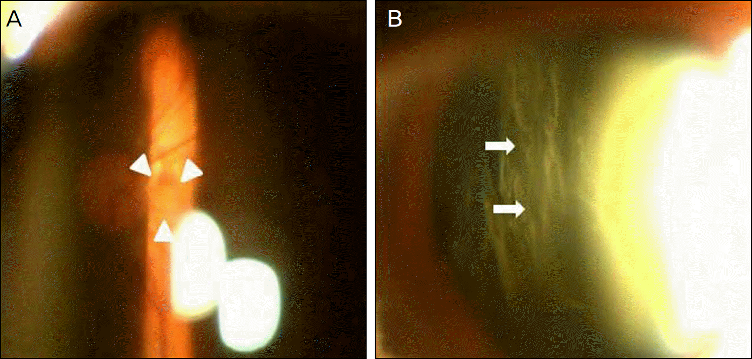
Figure 5.
The distribution of vitreomacular interface status evaluated with TD OCT and SD OCT (p < 0.0001).
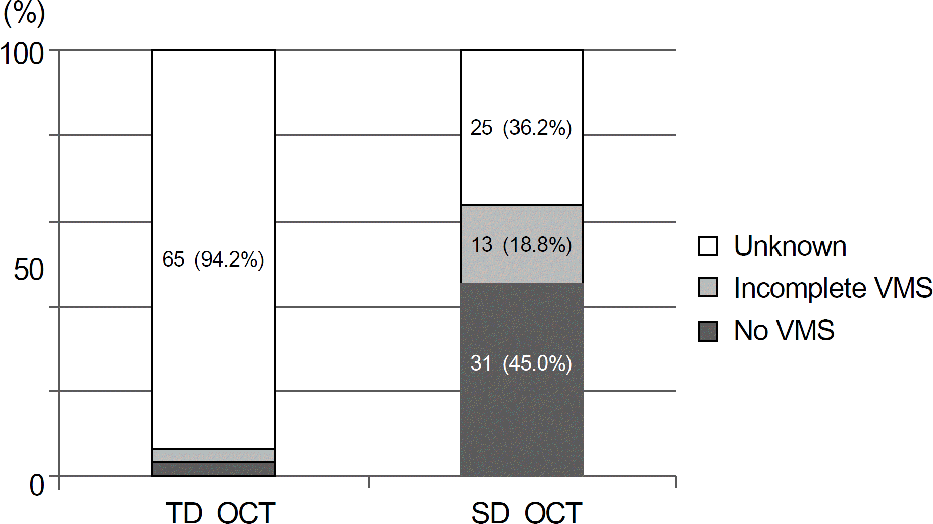
Figure 6.
Comparison of the efficacy of TD OCT and SD OCT in the evaluation of vitreomacular interface. Posterior hyaloid interface was visible in only 2 eyes with TD OCT of 31 eyes for which no VMS was identified on SD OCT (A, A1, A2). In 13 eyes with incomplete VMS on SD OCT, vitreomacular interfaces were visualized in only 2 eyes on TD OCT (B, B1, B2).
 XML Download