Abstract
Purpose
To report a case of anterior chamber intraocular lens (ACL) reposition with the haptic protruded into the subcon-junctiva in a patient with a previous ACL implantation.
Case summary
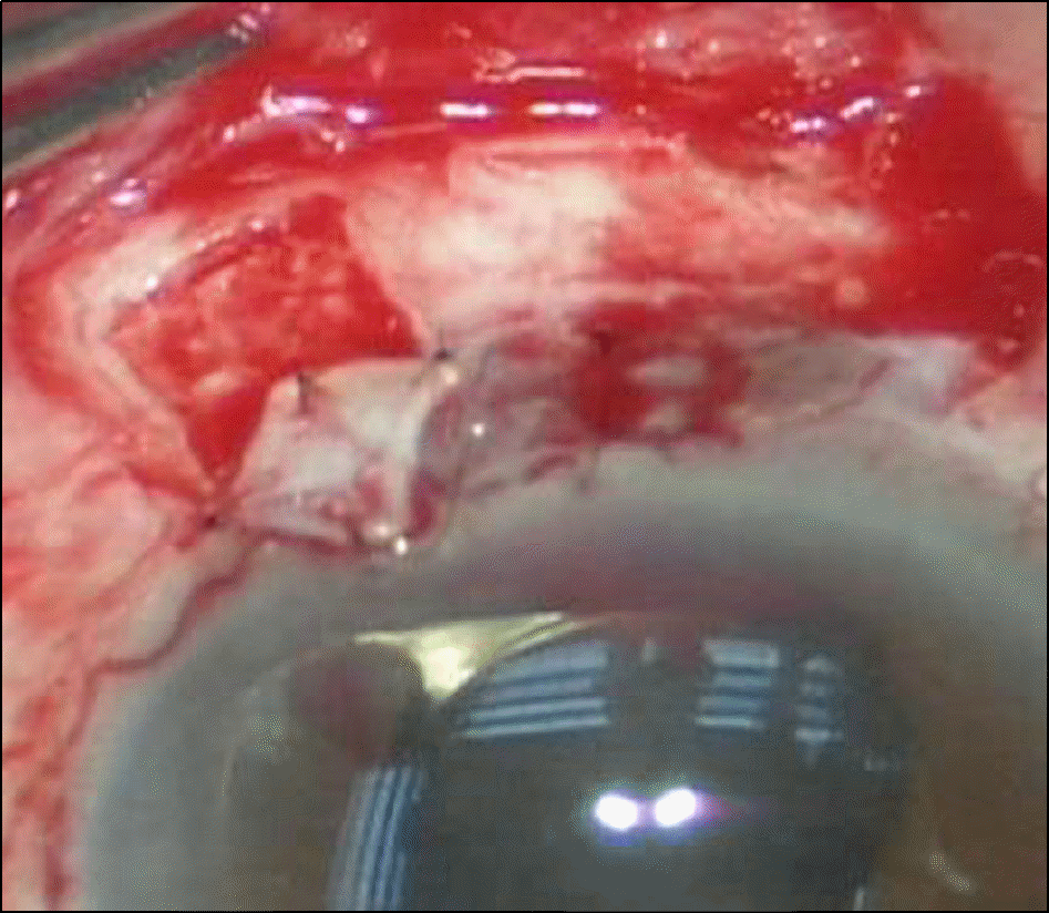
A 64-year-old man visited our clinic because of visual disturbance and discomfort in his right eye. Approximately 8 years earlier, he had cataract surgery and there was no visual improvement but eye discomfort. The hap-tic of the ACL protruded into the subconjunctiva at 11-1 o’clock. The visual acuity of the right eye was 0.2 and the intra-ocular pressure of the right eye was 27 mmHg. The ACL was repositioned because of low cell density (1222 cells/mm2). After 6 months, the visual acuity of the right eye was 0.3, best corrected visual acuity was 0.8, intraocular pressure was 12 mmHg and cell density was 838 cells/mm2. There were no inflammation signs or complications.
Go to : 
References
1. Kim SH, Kim IC, Chung YT. Results of Nuvita phakic anterior chamber intraocular lens for high myopia. J Korean Ophthalmol Soc. 2001; 42:1721–8.
2. Yi TK, Kwon JS, Shin DE. Visual outcome and complications of Modern Kelman style anterior chamber intraocular lens implantation. J Korean Ophthalmol Soc. 1999; 40:2773–84.
4. Yeom IY, Chang JH, Jung YC. A clinical study on implantation of anterior chamber intraocular lens and posterior chamber intra-ocular lens by scleral fixation in eyes without capsular or zonular support. J Korean Ophthalmol Soc. 1993; 34:950–5.
5. Jang BH, Lee DW, Cho NC, Ahn M. Clinical results of anterior chamber phakic intraocular lens. J Korean Ophthalmol Soc. 2006; 47:31–6.
6. Drews RC. The Barraquer experience with intraocular lenses. 20 years later. Ophthalmology. 1982; 89:386–93.

7. Wolter JR. Foreign body giant cells selectively covering haptics of intraocular lens implants. indicators of poor toleration? Ophthalmic Surg. 1983; 14:839–44.
8. Yeo JH, Jakobiec FA, Pokorny K. . The ultrastructure of an IOL “cocoon membrane”. Ophthalmology. 1983; 90:410–9.

9. Lee E, Park SJ, Kwon JD. A case of Ahmed glaucoma valve im-plantation without removal of the anterior chamber lens. J Korean Ophthalmol Soc. 2011; 52:746–52.

10. Kim SI, Na KS, Kwon HG. . Effect of anterior chamber depth on corneal endothelial change after phacoemulsification. J Korean Ophthalmol Soc. 2010; 51:1568–72.

11. Choi DW, Kim EJ, Chung IY. . Clinical study on anterior chamber intraocular lens implantation in completely vitrectom-ized eyes. J Korean Ophthalmol Soc. 2005; 46:45–50.
12. Oh SY, Lee JH. The effect of the posterior chamber IOL im-plantation by suture fixation of the number of central corneal endo-thelial cells. J Korean Ophthalmol Soc. 1994; 35:58–62.
Go to : 
 | Figure 1.Haptic of anterior chamber intraocular lens pro-trudes into subconjunctival space at 11-1 o’clock perilimbal area, and two endings of haptic expose (arrows). |
 | Figure 3.Anterior chamber intraocular lens is repositioned at 2-8 o’clock. Corneo-scleral hole is shown at the site where haptic was incarcerated previously (arrow). |


 PDF
PDF ePub
ePub Citation
 Print
Print


 XML Download