Abstract
Case summary
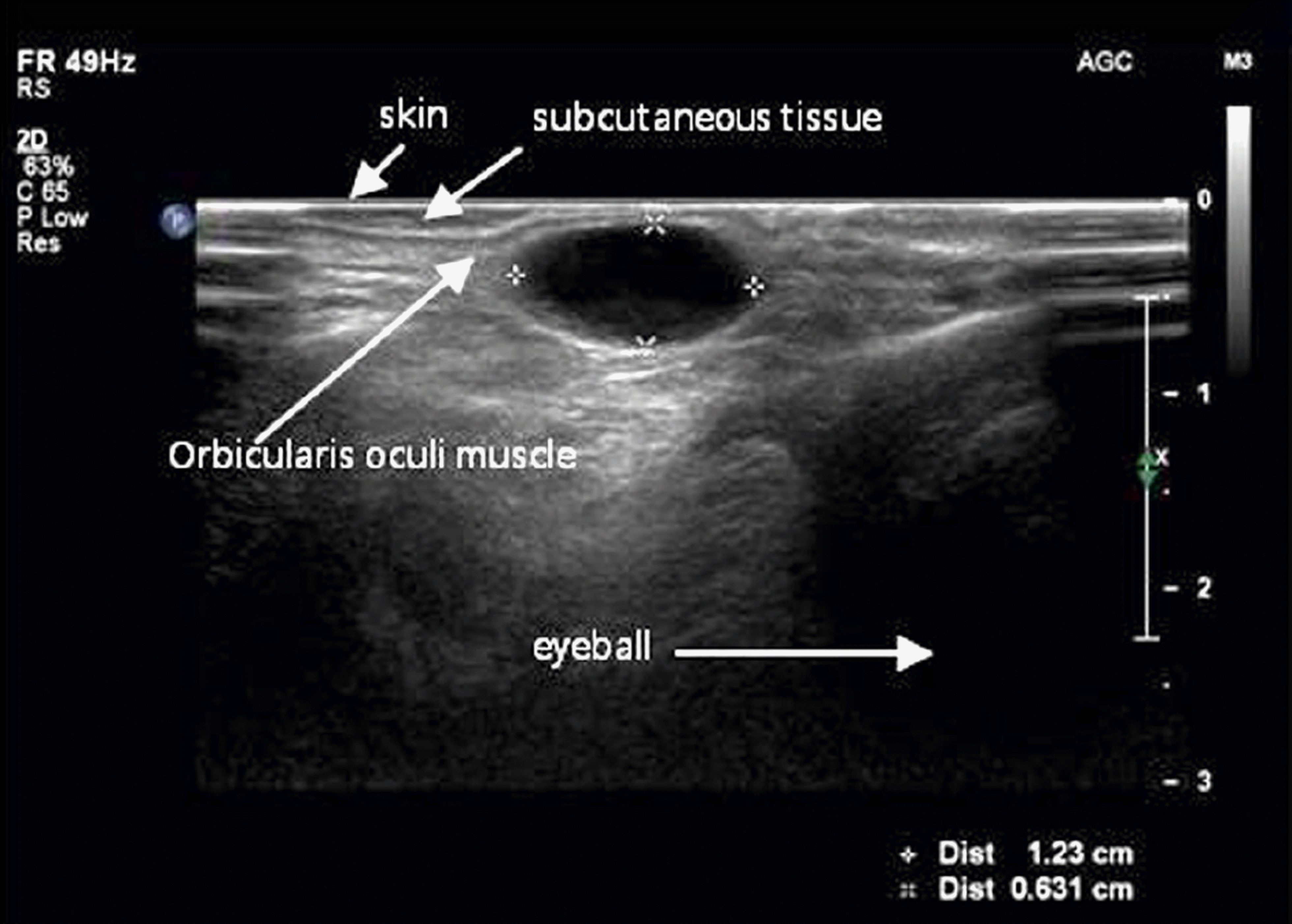
A 52-year-old male visited our clinic with an orbital mass in the right eye, which had developed 4 months prior to admission. A 2 × 1.5 cm-sized hard orbital mass was palpated in the middle area of the right upper orbit. On sono-graphic imaging, a well-demarcated tumor was identified that showed no echogenicity. We performed anterior orbitotomy with a lid crease incision. The tumor was completely removed. Histopathological examination showed a solitary cyst lined with two or three layers of cuboidal epithelial cells with scattered goblet cells. The tumor was classified as a sudoriferous cyst.
Go to : 
References
1. Wiliam HS. Eyelids and Lacrimal Drainage System. In: Jurij RB, ed. Ophthalmic Pathology. 4th ed.Philadelphia: WB Saunders;1996; v. 4:chap. 11.
2. Haider E, Saigal G, Gill D. . Congenital orbital sudoriferous cyst: radiological findings. Pediatr Radiol. 2005; 35:1142–4.

3. Kim JY, Lee EK, Lee YH, Lee SB. Apocrine sudoriferous cyst in medial canthal region occurred after trauma. J Korean Ophthalmol Soc. 2007; 48:1562–6.

4. Smith RJ, Kuo IC, Reviglio VE. Multiple apocrine hidrocystomas of the eyelids. Orbit. 2012; 31:140–2.

5. Mehta A, Rao A, Khanna A. Sudoriferous cyst of the orbit of adult origin after trauma. Indian J Ophthalmol. 2008; 56:235–7.

6. Chung JK, Lee SJ, Kang SK, Park SH. Congenital sudoriferous cyst within the orbit followed by esotropia. Korean J Ophthalmol. 2007; 21:120–3.

10. Park SJ, Jang JW, Chin HS. Sudoriferous cyst of the orbit. J Korean Ophthalmol Soc. 1998; 39:1288–90.
11. Mims J, Rodrigues M, Calhoun J. Sudoriferous cyst of the orbit. Can J Ophthalmol. 1977; 12:155–6.
Go to : 
 | Figure 1.Preoperative photograph showing right superomedial orbital swelling, fixed to the deep orbital planes on palpation. |
 | Figure 2.The ultrasound image of the right superomedial orbit showing the well-demarcated, thin walled, anechoic mass, consistent with cyst beneath the muscle layer. |
 | Figure 3.Hispathologic photographs of the specimen. (A) Low power view of the cyst showing areas of ductal cyst with fibrous tis-sue wall (H&E stain, ×40). (B) High power view of the cyst showing areas of double layered cuboidal cell lining, the inner layer showing apical snouts (H&E stain, ×400). (C) High power view of the cyst showing Periodic acid Schiff (PAS) positive apical gly-cocalyx (PAS stain, ×400). |


 PDF
PDF ePub
ePub Citation
 Print
Print


 XML Download