

 PDF
PDF ePub
ePub Citation
 Print
Print


Abstract
Purpose
To compare the mean deviation (MD) between monocular and integrated binocular visual field (BVF).
Methods
Thirty-six patients with glaucoma in at least 1 eye were recruited for the present study. Seventy-two threshold sensitivities of the BVF were obtained without additional visual field test by the Best Location and Binocular Summation methods using the 2 monocular visual fields of central 30 o. The MD of the BVF was obtained by comparison to the value distribution in the age-matched population with normal BVF. After defining the better eye with the better MD value from the 2 eyes, comparison of the MDs between individual eyes and the integrated BVF were assessed. In addition, the MDs be-tween the integrated BVF and actual BVF were compared in 11 patients.
Results
In patients with a mean age of 58.7 years, the MD of the better eye was -2.3 dB, and the worse eye was -4.9 dB (p < 0.01). There was a significant difference between the 2 MDs derived from Best Location and Binocular Summation (-1.7 and -2.0 dB, respectively p = 0.045). The MDs according to BVF more improved than the better eye (p < 0.01 for both). There was no significant difference in MDs between integrated BVF and actual BVF (-1.9 vs -2.0, -2.3 vs -2.0, re-spectively p > 0.1).
References
1. Kass MA, Heuer DK, Higginbotham EJ. . The Ocular Hypertension Treatment Study: a randomized trial determines that topical ocular hypotensive medication delays or prevents the onset of primary open-angle glaucoma. Arch Ophthalmol. 2002; 120:701–13.
2. Miglior S, Zeyen T, Pfeiffer N. . Results of the European Glaucoma Prevention Study. Ophthalmology. 2005; 112:366–75.
3. Gupta N, Krishnadev N, Hamstra SJ, Yücel YH. Depth perception deficits in glaucoma suspects. Br J Ophthalmol. 2006; 90:979–81.

4. Ramulu PY, West SK, Munoz, B. . Driving cessation and driving limitation in glaucoma: the Salisbury Eye Evaluation Project. Ophthalmology. 2009; 116:1846–53.
5. McGwin G Jr, Xie A, Mays A. . Visual field defects and the risk of motor vehicle collisions among patients with glaucoma. Invest Ophthalmol Vis Sci. 2005; 46:4437–41.
6. Patino CM, McKean-Cowdin R, Azen SP. . Central and periph-eral visual impairment and the risk of falls and falls with injury. Ophthalmology. 2010; 117:199–206.
7. Gutierrez P, Wilson MR, Johnson C. . Influence of glaucoma-tous visual field loss on health-related quality of life. Arch Ophthalmol. 1997; 115:777–84.
8. Janz NK, Wren PA, Lichter PR. . Quality of life in newly diag-nosed glaucoma patients : The Collaborative Initial Glaucoma Treatment Study. Ophthalmology. 2001; 108:887–97.
9. McKean-Cowdin R, Wang Y, Wu J. . Impact of visual field loss on health-related quality of life in glaucoma: the Los Angeles Latino Eye Study. Ophthalmology. 2008; 115:941–8.e1.
10. Parrish RK 2nd, Gedde SJ, Scott IU. . Visual function and quality of life among patients with glaucoma. Arch Ophthalmol. 1997; 115:1447–55.
11. van Gestel A, Webers CA, Beckers HJ. . The relationship be-tween visual field loss in glaucoma and health-related qual-ity-of-life. Eye (Lond). 2010; 24:1759–69.
12. Varma R, Wu J, Chong K. . Impact of severity and bilaterality of visual impairment on health-related quality of life. Ophthalmology. 2006; 113:1846–53.
13. Friedman DS, Freeman E, Munoz B. . Glaucoma and mobility performance: the Salisbury Eye Evaluation Project. Ophthalmology. 2007; 114:2232–7.
14. Owen VM, Crabb DP, White ET. . Glaucoma and fitness to drive: using binocular visual fields to predict a milestone to blindness. Invest Ophthalmol Vis Sci. 2008; 49:2449–55.
15. Jampel HD, Friedman DS, Quigley H, Miller R. Correlation of the binocular visual field with patient assessment of vision. Invest Ophthalmol Vis Sci. 2002; 43:1059–67.
16. Crabb DP, Viswanathan AC, McNaught AI. . Simulating bin-ocular visual field status in glaucoma. Br J Ophthalmol. 1998; 82:1236–41.
17. Nelson-Quigg JM, Cello K, Johnson CA. Predicting binocular vis-ual field sensitivity from monocular visual field results. Invest Ophthalmol Vis Sci. 2000; 41:2212–21.
18. Crabb DP, Fitzke FW, Hitchings RA, Viswanathan AC. A practical approach to measuring the visual field component of fitness to drive. Br J Ophthalmol. 2004; 88:1191–6.
19. Legge GE. Binocular contrast summation–II. Quadratic summation. Vision Res. 1984; 24:385–94.
20. Anderson DR. The single field printout with Statpac analysis. Kist K, ed. Automated Static Perimetry. St. Louis: MO Mosby;1992; 84.
21. Esterman B. Functional scoring of the binocular field. Ophthalmology. 1982; 89:1226–34.
22. Jampel HD. Glaucoma patients' assessment of their visual function and quality of life. Trans Am Ophthalmol Soc. 2001; 99:301–17.
23. Mills RP. Correlation of quality of life with clinical symptoms and signs at the time of glaucoma diagnosis. Trans Am Ophthalmol Soc. 1998; 96:753–812.
24. Campbell FW, Green DG. Monocular versus binocular visual acuity. Nature. 1965; 208:191–2.
25. Meese TS, Georgeson MA, Baker DH. Binocular contrast vision at and above threshold. J Vis. 2006; 6:1224–43.
26. Simpson WA, Manahilov V, Shahani U. Two eyes: square root 2 better than one? Acta Psychol (Amst). 2009; 131:93–8.
27. Asaoka R, Crabb DP, Yamashita T. . Patients have two eyes!: binocular versus better eye visual field indices. Invest Ophthalmol Vis Sci. 2011; 52:7007–11.
Figure 1.
Schematic representation of the left (A), right (B), in-tegrated binocular visual field (BVF) by best location (D), BVF by binocular summation (E), and actual binocular visual field test (E). Four points (black squares of BVF) corresponding to the blind spots of both eyes without correlation between the right eye and left eye were excluded and threshold sensitivities of BVF were integrated from right and left eyes. VF (visual field).
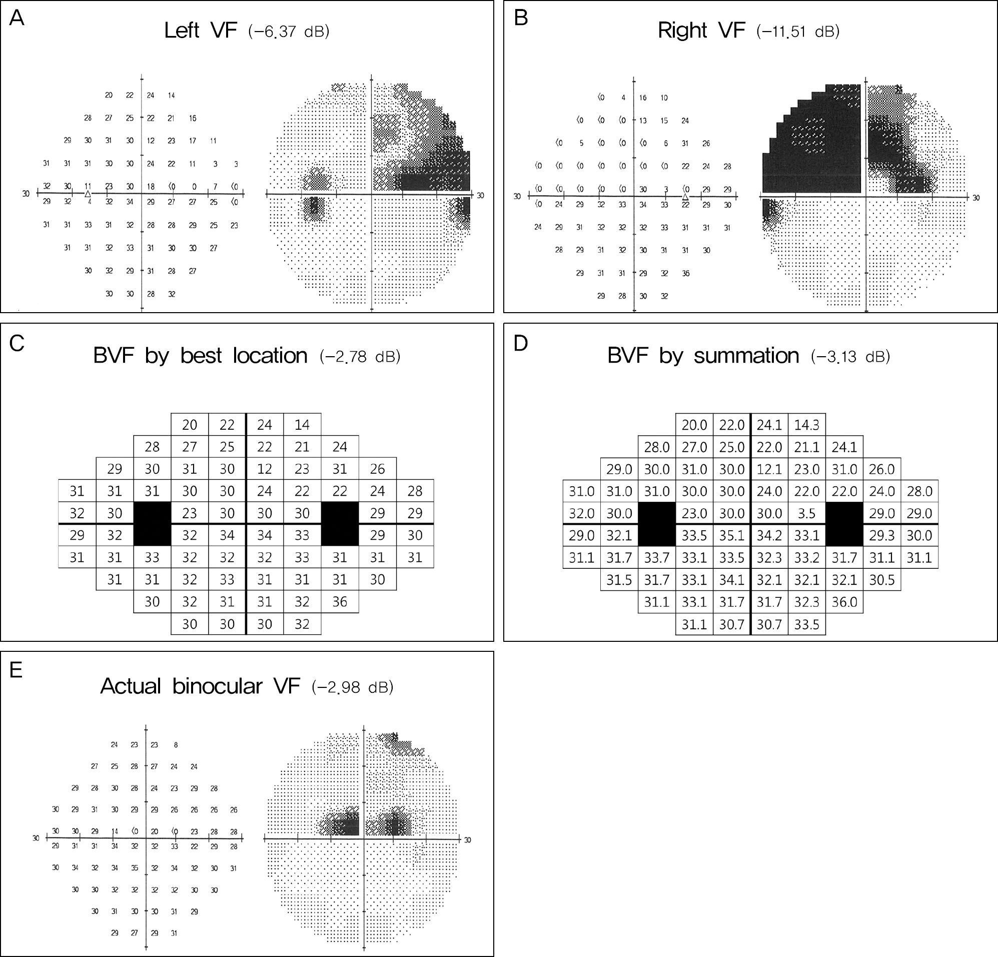
Figure 2.
Boxplot for the mean deviations of the integrated binocular visual field (BVF) by best location and binocular summation, the better and worse eye visual fields.
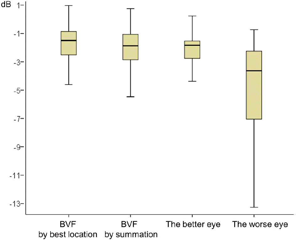
Table 1.
Demographic and clinical characteristics of the patient and control groups in the study
| Patient group | Control group | p-value* | |
|---|---|---|---|
| Number of included subjects | 36 | 30 | |
| Age (year) | 58.7 ± 8.9 | 59.3 ± 10.2 | 0.194 |
| Sex (F/M) (n (%)) | 19 (52.8)/17 (47.2) | 17 (56.7)/13 (43.3) | 0.282 |
| IOP (mm Hg)† | 14.8 ± 3.8 | 15.0 ± 2.5 | 0.420 |
| Follow-up (year) | 3.8 ± 1.3 | 1.2 ± 0.4 | <0.01 |
| Total number of VF (pairs) | 72 (36) | 60 (30) | |
| MD of right VF, dB‡ | -4.4 ± 3.5 (-5.9, -3.1, -1.9) | -0.3 ± 1.3 (-1.1, -0.3, 0.7) | <0.01 |
| MD of left VF, dB‡ | -2.9 ± 2.4 (-4.1, -1.9, -1.7) | -0.4 ± 1.5 (-1.3, -0.6, 1.0) | <0.01 |
Table 2.
Mean deviation on visual field of the patient group (n = 36)
| Mean deviation, dB* | p-value† | p-value‡ | |
|---|---|---|---|
| Right VFs | -4.4 ± 3.5 (-5.9, -3.1, -1.9) | <0.01 | <0.01 |
| Left VFs | -2.9 ± 2.4 (-4.1, -1.9, -1.7) | <0.01 | <0.01 |
| BVF by best location | -1.7 ± 1.4 (-2.5, -1.5, -0.9) | 0.045 | |
| BVF by binocular summation | -2.0 ± 1.6 (-2.9, -1.9, -1.0) | 0.045 | |
| The better eye | -2.3 ± 2.0 (-3.0, -1.9, -1.5) | 0.035 | <0.01 |
| The worse eye | -4.9 ± 3.4 (-7.0, -3.6, -2.2) | <0.01 | <0.01 |
Table 3.
Comparison of mean deviation between integrated BVF and actual BVF in the 11 patients
| Mean deviation, dB* | p-value† | |
|---|---|---|
| Actual BVF | -2.0 ± 2.7 (-4.5, -2.0, 1.0) | |
| BVF by best location | -1.9 ± 2.4 (-4.3, -2.1, 0.4) | 0.858 |
| BVF by binocular summation | -2.3 ± 2.7 (-5.1, -2.5, 0.3) | 0.172 |
Table 4.
Mean deviation on visual field of the patient with glaucoma at both eyes or patients with glaucoma at only one eye
| Mean deviation, dB* | p-value† | p-value‡ | |
|---|---|---|---|
| Patients with glaucoma at both eyes (n = 14) | |||
| BVF by best location | -2.3 ± 1.6 (-3.5, -2.1, -1.1) | <0.01 | |
| BVF by binocular summation | -2.7 ± 1.7 (-3.7, -2.4, -1.6) | <0.01 | |
| The better eye | -3.7 ± 2.4 (-5.6, -3.4, -1.9) | 0.036 | 0.041 |
| The worse eye | -6.7 ± 3.6 (-8.8, -5.9, -3.5) | <0.01 | <0.01 |
| Patients with glaucoma at only one eye (n = 22) | |||
| BVF by best location | -1.3 ± 1.3 (-1.9, -1.2, -0.7) | 0.038 | |
| BVF by binocular summation | -1.4 ± 1.1 (-1.9, -1.7, -0.4) | 0.038 | |
| The better eye | -1.5 ± 1.3 (-2.2, -1.2, -0.8) | <0.01 | 0.096 |
| The worse eye | -3.8 ± 2.7 (-5.1, -2.6, -2.0) | <0.01 | <0.01 |
 XML Download