Abstract
Purpose
To evaluate if the presence of pathologic myopia could affect the result of macular hole surgery.
Methods
This study was a retrospective comparison of the results of macular hole surgery between a pathologic myopia group (11 eyes) and a non-pathologic myopia group (14 eyes). All patients had undergone PPV, ILM peeling and C3F8 (20%) gas temponade. BCVA, IOP and OCT findings were evaluated preoperatively and at 6 months after surgery. Postoperative BCVA, IOP and macular hole closure were compared between each groups.
Results
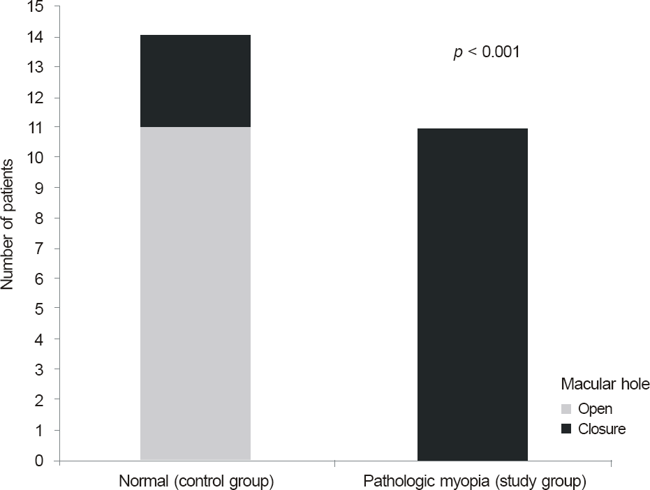
The only statistically significant preoperative parameter between the groups was axial length (p < 0.001). Postoperative BCVA was lower in the pathologic myopia group, but the difference was not statistically significant. The rate of macular hole closure was statistically significant higher in the non-pathologic myopia group (p < 0.001).
Go to : 
References
1. Kelly NE, Wendel RT. Vitreous surgery for idiopathic macular holes: Results of a pilot study. Arch Ophthalmol. 1991; 109:654–9.

2. Freeman WR, Azen SP, Kim JW. . Vitrectomy for the treatment of full-thickness stage 3 or 4 macular holes. Results of a multi-centered randomized clinical trial. The Vitrectomy for Treatment of Macular Hole Study Group. Arch Ophthalmol. 1997; 115:11–21.

3. Brooks HL Jr.Macular hole surgery with and without internal lim-iting membrane peeling. Ophthalmology. 2000; 107:1939–48.

4. Kang HK, Chang AA, Beaumont PE. The macular hole: report of an Australian surgical series and meta-analysis of the literature. Clin Experiment Ophthalmol. 2000; 28:293–308.

5. Park DW, Lee JH, Min WK. The use of internal limiting membrane maculorrhexis in treatment of idiopathic macular holes. Korean J Ophthalmol. 1998; 12:92–7.

6. Smiddy WE, Feuer W, Cordahi G. Internal limiting membrane peeling in macular hole surgery. Ophthalmology. 2001; 108:1471–6.

7. Haritoglou C, Gass CA, Schaumberger M. . Long-term fol-low-up after macular hole surgery with internal limiting membrane peeling. Am J Ophthalmol. 2002; 134:661–6.

8. Xirou T, Xirou V, Mangouritsas G. . Full thickness macular hole closure after exchanging silicone-oil tamponade with C(3)F(8) without posturing. Case Report Ophthalmol. 2011; 2:166–9.

9. Nishimura A, Kimura M, Saito Y, Sugiyama K. Efficacy of pri-mary silicone oil tamponade for the treatment of retinal detach-ment caused by macular hole in high myopia. Am J Ophthalmol. 2011; 151:148–55.

10. Tafoya ME, Lambert HM, Vu L, Ding M. Visual outcomes of sili-cone oil versus gas tamponade for macular hole surgery. Semin Ophthalmol. 2003; 18:127–31.

11. Lai JC, Stinnett SS, McCuen BW. Comparison of silicone oil ver-sus gas tamponade in the treatment of idiopathic full-thickness macular hole. Ophthalmology. 2003; 110:1170–4.

12. Qu J, Zhao M, Jiang Y, Li X. Vitrectomy outcomes in eyes with high myopic macular hole without retinal detachment. Retina. 2012; 32:275–80.

13. Wu TT, Kung YH. Comparison of anatomical and visual outcomes of macular hole surgery in patients with high myopia vs. non-high myopia: a case-control study using optical coherence tomography. Graefes Arch Clin Exp Ophthalmol. 2012; 250:327–31.
14. Ikuno Y, Tano Y. Vitrectomy for macular holes associated with my-opic foveoschisis. Am J Ophthalmol. 2006; 141:774–6.

15. Scholda C, Wirtitsch M, Biowski R, Stur M. Primary silicone oil tamponade without retinopexy in highly myopic eyes with central macular hole detachments. Retina. 2005; 25:141–6.

16. Grossniklaus HE, Green WR. Pathologic finding in pathologic myopia. Retina. 1992; 12:127–33.
17. Oster SF, Mojana F, Bartsch DU. . Dynamics of the macular hole-silicone oil tamponade interface with patient positioning as imaged by spectral domain-optical coherence tomography. Retina. 2010; 30:924–9.

18. Feng LG, Jin XH, Li JK. . Surgical management of retinal de-tachment resulting from macular hole in a setting of high myopia. Yan Ke Xue Bao. 2012; 27:69–75.
19. Nadal J, Verdaguer P, Canut MI. Treatment of retinal detachment secondary to macular hole in high myopia: Vitrectomy with dis-section of the inner limiting membrane to the edge of the staph-yloma and long-term tamponade. Retina. 2012; 32:1525–30.
Go to : 
 | Figure 1.Comparison of postoperative 6-month macular hole closure between groups. Macular hole closure was observed in 11 patients after primary surgery in control group (idiopathic macular hole group). In contrast, any macular hole closure was not observed in study group (pathologic myopia group). |
Table 1.
Baseline characteristic of study group (pathologic myopia group)
Table 2.
Baseline characteristic of control group
Table 3.
Comparison of outcomes between the study group and control group
| High myopic MH Group (Study group) | Idiopathic MH Group (Control group) | p-value | |
|---|---|---|---|
| Eye | 11 | 14 | |
| Age (years) | 61.45 ± 10.41 | 67.57 ± 7.89 | 0.317 |
| Diabetes | 7/11 (63.6%) | 7/14 (50%) | 0.689 |
| Macular hole size (μ m) | 733.36 ± 286.89 | 643.43 ± 296.00 | 0.406 |
| Axial length (mm) | 30.64 ± 1.78 | 22.93 ± 1.02 | <0.001† |
| Preop VA (log MAR) | 0.69 ± 0.29 | 0.89 ± 0.28 | 0.085 |
| Postop VA (log MAR) | 0.80 ± 0.34 | 0.50 ± 0.44 | 0.075 |
| Preop IOP (mm Hg) | 15.45 ± 1.57 | 14.93 ± 4.16 | 0.572 |
| Postop IOP (mm Hg) | 15.64 ± 2.84 | 14.57 ± 3.08 | 0.291 |
| IOP elevation history | 4/11 (36.4%) | 1/14 (7.1%) | 0.070 |
| Macular hole closure (%) | 0/11 (0%) | 11/14 (78.6%) | <0.001† |
| Retreatment success | 3 (gas) → fail | 3 (gas) → sucess | |
| 2 (silicone oil) → sucess |


 PDF
PDF ePub
ePub Citation
 Print
Print


 XML Download