Abstract
Purpose
To describe the clinical manifestations, radiologic findings, and treatment outcomes of pediatric pseudotumors. Methods: A retrospective chart review of patients diagnosed with pediatric pseudotumor (age under 20 years old) from August 2008 to February 2012 was performed.
Results
Thirteen patients (16 eyes) were included in this study. The mean age of the subjects was 14.2 years (5-20 years). Swollen eyelid (56.3%), ptosis (43.8%), conjunctival injection (18.8%), localized mass (18.8%), limitation of ocular move-ment (12.5%), proptosis (6.3%), and decreased visual acuity (6.3%) were found initially. Dacryoadenitis (62.5%), myositis (18.8%), anterior orbital inflammation (18.8%), and diffuse type (6.3%) were observed on orbital computed tomography (CT). Among the 13 patients (16 eyes), 8 patients (10 eyes) were administered oral systemic corticosteroids, 2 patients (2 eyes) received IV systemic corticosteroid, 1 patient (1 eye) received systemic corticosteroids combined with NSAID, and 2 patients (3 eyes) were prescribed NSAIDs only. Symptoms improved 4.1 days after initiation of treatment. CT scans re-vealed that one patient experienced diffuse-type disease recurrence twice.
Go to : 
References
1. Yan J, Qui H, Wu Z, Li Y. Idiopathic orbital inflammatory pseudo-tumor in Chinese children. Orbit. 2006; 25:1–4.

2. Mulvihill A, Smith CR, Buncic JR. Pediatric orbital pseudotumor presenting as a painless orbital and periocular mass. Can J Ophthalmol. 2004; 39:77–9.
3. Yuen KS, Lai CH, Chan WM, Lam DS. Bilateral exudative retinal attachdements as the presenting features of idiopathic orbital inflammation. Clin Experiment Ophthalmol. 2005; 33:671–4.
4. Mottow LS, Jakobiec FA. Idiopathic inflammatory orbital pseudo-tumor in childhood. I. Clinical characteristics. Arch Ophthalmol. 1978; 96:1410–7.

5. Nugent RA, Rootman J, Robertson WD. . Acute orbital pseu-dotumors, classification and CT features. AJR Am J Roentgenol. 1981; 137:957–62.

6. Chung SA, Yoon JS, Lee SY. Effect of intravenous methyl-prednisolone on idiopathic orbital inflammation. J Korean Ophthalmol Soc. 2010; 51:1299–304.

7. Berger JW, Rubin PA, Jakobiec FA. Pediatric orbital pseudotumor: Case report and review of the literature. Int Ophthalmol Clin. 1996; 36:161–77.

8. Stevens JL, Rychwalski PJ, Baker RS, Kielar RS. Pseudotumor of the orbit in early childhood. J AAPOS. 1998; 2:120–3.

9. Park SJ, Sin SJ, Lee DG, Jang JW. Pseudotumor: distribution, clin-ical features, treatment outcomes. J Korean Ophthalmol Soc. 2008; 49:1379–86.
10. Mombaerts I, Koornneef L. Current status in the treatment of orbi-tal myositis. Ophthalmology. 1997; 104:402–8.

11. Mombaerts I, Schlingemann RO, Goldschmeding R, Koornneef L. Are systemic corticosteroids useful in the management of orbital pseudotumors? Ophthalmology. 1996; 103:521–8.

12. Jordan DR. Orbital pseudotumors treated with systemic corticosteroids. Ophthalmology. 1996; 103:1519.

13. Yan J, Wu Z, Li Y. The differentiation of idiopathic inflammatory pseudotumor from lymphoid tumors of orbit: analysis of 319 cases. Orbit. 2004; 23:245–54.

14. Lee H, Kim SJ, Lee SY. Classification and treatment efficacy of orbital pseudotumor. J Korean Ophthalmol Soc. 2001; 42:1647–54.
15. Espinoza GM. Orbital inflammatory pseudotumors: etiology, dif-ferential diagnosis, and management. Curr Rheumatol Rep. 2010; 12:443–7.

16. Yuen SJ, Rubin PA. Idiopathic orbital inflammation: distribution, clinical features, and treatment outcome. Arch Ophthalmol. 2003; 121:491–9.
Go to : 
 | Figure 1.Case 4. Fundus photography of the patient's left eye. The patient presented with decreased vision of left eye. Note the disc swelling and retinal folds, supporting the diagnosis of posterior perineuritis. |
 | Figure 2.Case 4. MRI coronal (A) and axial (B) images of T1 gadolinium-enhanced slice at day of presentation. Inflammatory mass is evident within the orbital apex with associated proptosis of the left eye. |
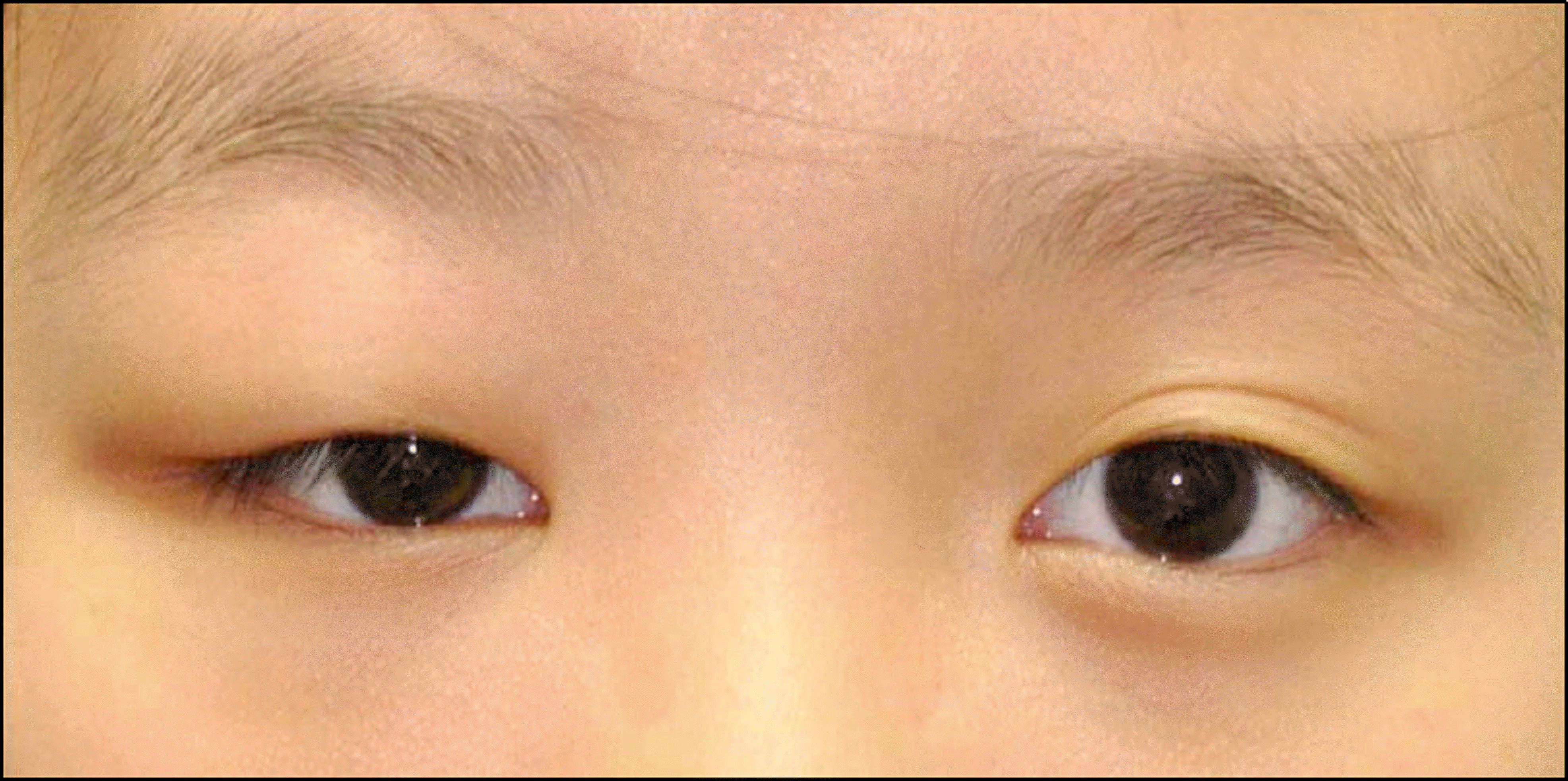
 | Figure 3.External photographs of case 6. Case 6 presented with non-erythematous swelling of right upper eyelid. The patients whose CT scan showed myositis involved the superior rectus muscle with improving ocular symptoms after receiving NSAID treatment. |
 | Figure 4.External photograph (A) and CT scan (B) of case 7. Case 7 presented with erythematous swelling of the left upper and lower eyelids. (A) CT scan shows left lacrimal gland en-largement and marked soft tissue thickening around the peri-orbit with dirty density being compatible with dacryoadenitis and periorbital cellulitis. (B) The patient received ex-perimental antibiotic treatment, however ocular symptoms did not improve. The patient showed improvement after receiving oral steroids. |
 | Figure 5.Dacryoadenitis (A). CT scan shows both lacrimal gland enlargement without focal lesion. Myositis (B). CT scan shows enlargement of the left inferior rectus muscle involving the tendon sheath. Anterior orbital inflammation (C). CT im-age shows ragged infiltrations involving the anterior soft tissue of the right orbit encompassing the right globe. |
Table 1.
Demographics and clinical features of the subjects


 PDF
PDF ePub
ePub Citation
 Print
Print


 XML Download