

 PDF
PDF ePub
ePub Citation
 Print
Print


Abstract
Case summary
A 22-year-old woman presented with a 1-year history of epiphora after two-jaw surgery. Orbital CT showed 5 mm of focal soft tissue at the level of the distal nasolacrimal duct. Dacryocystography showed complete ob-struction at the nasolacrimal duct level. Thus a nasolacrimal duct obstruction was diagnosed by physical and radiologic examination.
Go to : 
References
1. Menendez LF, Biedlingmaier JF, Tilghman D. Osteomeatal com-plex obstruction and sinusitis following Le Fort I osteotomy. J Oral Maxillofac Surg. 1996; 54:103–4.

2. Bruno C, Fernanda N, Belini M. Bloody tears after miniplate osteo-synthesis for Le Fort I osteotomy. Asian J Oral Maxillofac Surg. 2011; 10:1016–8.
3. Shoshani Y, Samet N, Ardekian L, Taicher S. Nasolacrimal duct in-jury after Le Fort I osteotomy. J Oral Maxillofac Surg. 1994; 52:406–7.
4. Keller EE, Sather AH. Quadriangular Le Fort I osteotomy : Surgical technique and review of 54 patients. J Oral Maxillofac Surg. 1990; 48:2–11.
5. Little C, Mintz S, Elinger AC. The distal lacrimal ductal system and traumatic epiphora. Int J Oral Maxillofac Surg. 1991; 20:31–5.
6. Demas PN, Sotereanos GC. Incidence of nasolacrimal injury and turbinectomy associated atrophic rhinitis with Le Fort I osteotomies. J Craniomaxillofac Surg. 1989; 17:116–8.
7. You ZH, Bell WH, Finn RA. Location of the nasolacrimal canal in relation to the high Le Fort I Osteotomy. J Oral Maxillofac Surg. 1992; 50:1075–80.
8. Bays RA, Bouloux GF. Complications of orthognathic surgery. Oral Maxillofacial Surg Clin N Am. 2003; 15:229–42.
9. Kim SG, Park SS. Incidence of complications and problems related to orthognathic surgery. J Oral Maxillofac Surg. 2007; 65:2438–44.
10. Li KK, Meara JG, Rubin PA. Orbital compartment syndrome fol-lowing orthognathic surgery. J Oral Maxillofac Surg. 1995; 53:964–8.
Go to :
 | Figure 3.A Dacryocystography of the patient shows complete obstruction of the right nasolacrimal duct. |
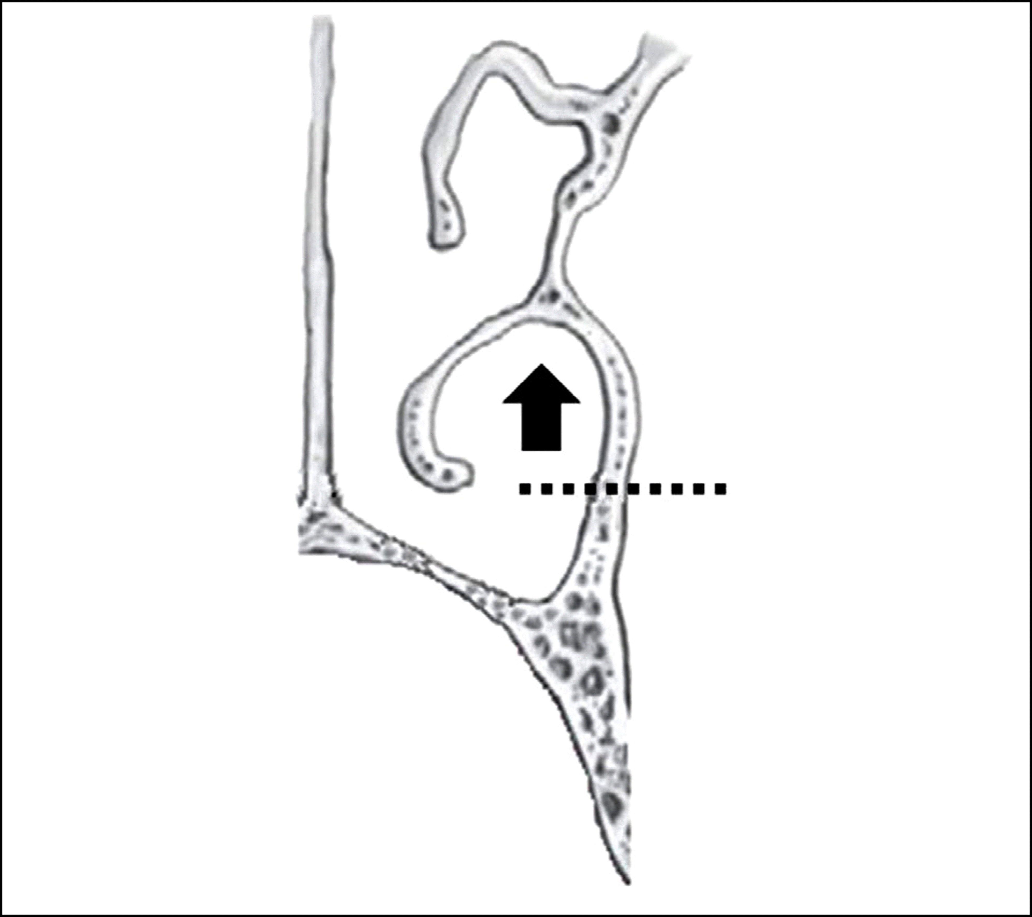
 | Figure 4.The osteotomy line of conventional Lefort surgery is shown by the dotted line. The inferior orifice of the nasolacri-mal canal (black arrow) is located at the top of the curved in-sertion of the inferior turbinate. |

 XML Download