

 PDF
PDF ePub
ePub Citation
 Print
Print


Abstract
Methods
Records of patients who underwent penetrating keratoplasty at the age of 5 years or younger were retro-spectively reviewed. The survival rates of corneal grafts, postoperative complications, and causes of graft failure were evaluated.
Results
A total of 31 penetrating keratoplasties were performed in 29 patients, two of which were bilateral. The mean fol-low-up period was 78.72 ± 8.94 months. The overall graft survival rate was 51.61%. The graft survival rate was 77.4% at 6 months, 61.3% at 12 months, 57.5% at 2 years, and 49.5% at 5 years after the surgery (the median survival time, 39.2 months). The main surgical indications included sclerocornea (35.5%), followed by Peter’s anomaly (25.8%) and congenital glaucoma (9.7%). There were significant differences in graft survival time among the surgical indications, of which scle-rocornea was the worst ( p = 0.003). The main cause of graft failure was rejection (46.7%), followed by infection (26.7%) and primary endothelial decompensation (20%). When patients were sub-grouped according to age (under 12 months, between 12 to 48 months, and over 48 months), there was significant difference in graft survival time ( p = 0.037) but not in overall graft survival rate ( p = 0.154). Graft rejection occurred more frequently in patients between 12 to 48 months of age compared to other age groups ( p = 0.016). Three out of 13 graft infections occurred in patients under 12 months of age.
Go to : 
References
1. Bermejo E, Martinez-Frias ML. Congenital eye malformations: clinical-epidemiological analysis of 1,124,654 consecutive births in Spain. Am J Med Genet. 1998; 75:497–504.

2. Ganekal S, Gangangouda C, Dorairaj S, Jhanji V. Early outcomes of primary pediatric keratoplasty in patients with acquired, atrau-matic corneal pathology. J AAPOS. 2011; 15:353–5.
3. Vanathi M, Panda A, Vengayil S, et al. Pediatric keratoplasty. Surv Ophthalmol. 2009; 54:245–71.
4. Al-Ghamdi A, Al-Rajhi A, Wagoner MD. Primary pediatric keratoplasty: indications, graft survival, and visual outcome. J AAPOS. 2007; 11:41–7.
5. McClellan K, Lai T, Grigg J, Billson F. Penetrating keratoplasty in children: visual and graft outcome. Br J Ophthalmol. 2003; 87:1212–4.
6. Stulting RD, Sumers KD, Cavanagh HD, et al. Penetrating kerato-plasty in children. Ophthalmology. 1984; 91:1222–30.
7. Lowe MT, Keane MC, Coster DJ, Williams KA. The outcome of corneal transplantation in infants, children, and adolescents. Ophthalmology. 2011; 118:492–7.
8. Dana MR, Schaumberg DA, Moyes AL, Gomes JA. Corneal trans-plantation in children with Peters anomaly and mesenchymal dysgenesis. Multicenter Pediatric Keratoplasty Study. Ophthalmology. 1997; 104:1580–6.
9. Rao KV, Fernandes M, Gangopadhyay N, et al. Outcome of pene-trating keratoplasty for Peters anomaly. Cornea. 2008; 27:749–53.
10. Yang LL, Lambert SR, Drews-Botsch C, Stulting RD. Long-term visual outcome of penetrating keratoplasty in infants and children with Peters anomaly. J AAPOS. 2009; 13:175–80.
11. Yang LL, Lambert SR, Lynn MJ, Stulting RD. Long-term results of corneal graft survival in infants and children with peters anomaly. Ophthalmology. 1999; 106:833–48.
12. Huang C, O'Hara M, Mannis MJ. Primary pediatric keratoplasty: indications and outcomes. Cornea. 2009; 28:1003–8.
Go to :
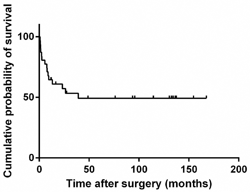 | Figure 1.The Kaplan-Meier survival curve of corneal grafts. The overall graft survival rate was 51.6%. The graft survival was 77.4% at 6 months, 61.3% at 12 months, 57.5% at 2 years and 49.5% at 5 years after the surgery. |
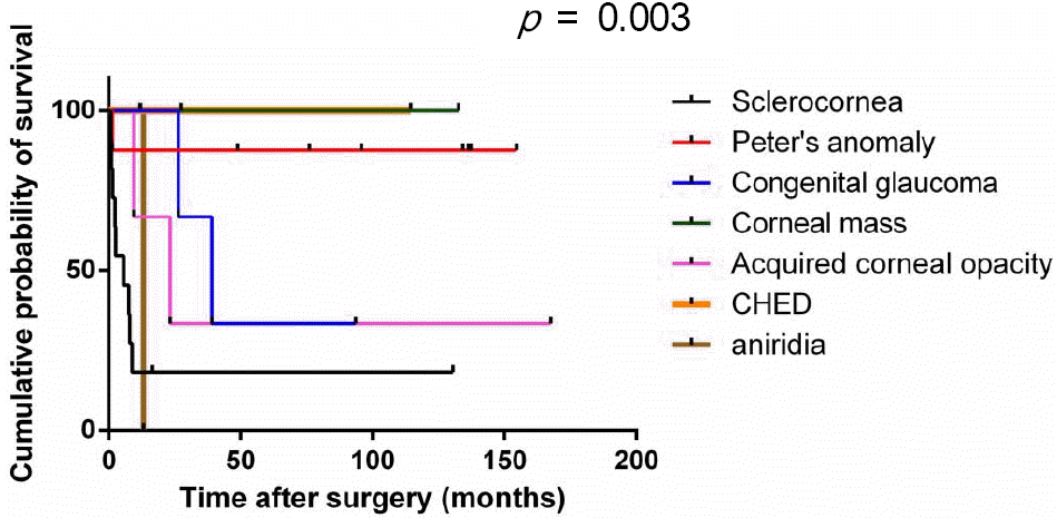 | Figure 2.The Kaplan-Meier survival curve of corneal grafts according to the surgical indications. Corneal mass, CHED and Peter’s anomaly show favorable outcome whereas scle-rocornea, congenital glaucoma, acquired corneal opacities and aniridia showed poor outcome. The differences of median sur-vival time among the surgical indications reached statistical significance ( p = 0.003, Log-Rank test). |
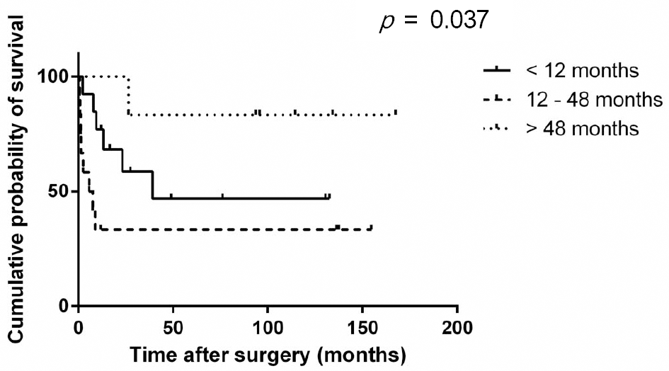 | Figure 3.The Kaplan-Meier survival curve of corneal grafts according to the age groups. The mean survival time of cor-neal grafts according to the age groups was significantly dif-ferent; 72.26 ± 17.62 months in age under 12 months vs 54.03 ± 20.57 months in age 12 to 48 months vs 144.03 ±14.60 months in age over 48 months ( p = 0.037, Log-Rank test). |
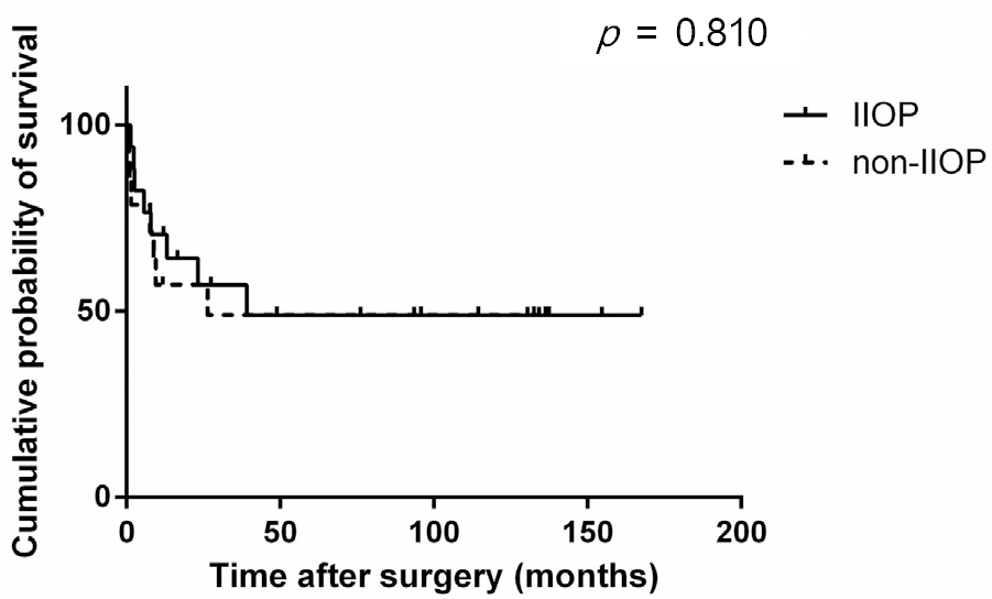 | Figure 4.The Kaplan-Meier survival curve of corneal grafts according to the use of topical intraocular pressure (IOP) low-ering agents. The median survival time was not significantly different between the patients who used IOP lowering agents due to increased IOP after the surgery and those who did not ( p = 0.810, Log-Rank test). |
Table 1.
Patient demographics
Table 2.
Complications after the surgery
| Complications | |
|---|---|
| Rejection | 45.2% (n = 14) |
| IIOP | 32.3% (n = 10) |
| Cataract | 25.8% (n = 8) |
| Phthisis / Enucleation | 22.6% (n = 7) |
| Infection | 16.1% (n = 5) |
| Causative agents* | Achromobacter xylosoxidans |
| Streptococcus pneumonia | |
| Herpes simplex virus |
Table 3.
Survival rates, rejection and infection rates according to age groups
| Age | <12 months | 12-48 months | >48 months | p-value* |
|---|---|---|---|---|
| N | 13 | 12 | 6 | |
| Survival | 53.85% (7/13) | 33.33% (4/12) | 83.33% (5/6) | 0.154 |
| Rejection | 7.69% (1/13) | 50% (6/12) | 0% | 0.016 |
| Infection | 23.08% (3/13) | 0% | 16.67% (1/6) | 0.220 |
Table 4.
The distribution of the final diagnosis according to the age groups
| Diagnosis | Age | N (%) | p-value* | ||
|---|---|---|---|---|---|
| <12 months | 12-48 months | >48 months | |||
| Sclerocornea | 4 (36%) | 7 (64%) | 0 (0%) | 11 (35.5%) | |
| Peter's anomaly | 2 (25%) | 4 (50%) | 2 (25%) | 8 (25.8%) | |
| Acquired cases | 2 (67%) | 0 (0%) | 1 (33%) | 3 (9.7%) | |
| Congenital glaucoma | 1 (33%) | 0 (0%) | 2 (67%) | 3 (9.7%) | 0.091 |
| Corneal mass | 3 (100%) | 0 (0%) | 0 (0%) | 3 (9.7%) | |
| CHED | 0 (0%) | 1 (50%) | 1 (50%) | 2 (6.5%) | |
| Aniridia | 1 (100%) | 0 (0%) | 0 (0%) | 1 (3.2%) |
 XML Download