

 PDF
PDF ePub
ePub Citation
 Print
Print


Abstract
Methods
In this prospective study, we determined the clinical features of pingueculae for 120 patients who visited our hos-pital due to pinguecula-related symptoms between October 2011 and May 2012.
Results
Among the 374 pingueculae of 120 patients, 208 (56%) pingueculae were found on the nasal side of the con-junctiva and 166 (44%) on the temporal side. The grade of the pingueculae on the temporal conjunctiva was 0.81 ± 0.626 and was 1.18 ± 0.648 on the nasal conjunctiva, which was higher ( p < 0.001). Pingueculae on the nasal conjunctiva showed greater elevation than on the temporal side ( p < 0.001). The horizontal and average diameters of the pingueculae on the nasal and temporal conjunctiva showed no significant difference, but the vertical diameters were 2.40 ± 0.88 mm and 2.14 ± 0.91 mm, respectively, showing the vertical diameter on the nasal conjunctiva was significantly greater ( p < 0.044). Moreover, the horizontal diameter of each pinguecula was greater than the vertical diameter ( p < 0.003).
Conclusions
The average grade of pingueculae in North-western Gyeonggi Province increased with age. Compared to the temporal pingueculae, the nasal pingueculae were more protrudent in shape and had higher grades. Furthermore, each pinguecula was oval-shaped with a larger horizontal than vertical diameter. The pingueculae on the temporal side were flatter and more oval-shaped.
Go to : 
References
1. Oh HJ, Park YG, Yoon KC. Changes of ocular surface and tear film in patients with pinguecula and pterygium. J Korean Ophthalmol Soc. 2006; 47:717–24.
2. Bell A. Pinguecula. J Vis Commun Med. 2006; 29:82–3.

3. Mimura T, Usui T, Obata H, et al. Severity and determinants of pin-guecula in a hospital-based population. Eye Contact Lens. 2011; 37:31–5.
4. Shin JY, Khang MH, Han YK, Kwon JW. Case of argon laser pho-toablation of pinguecula. Clin Experiment Ophthalmol. 2010; 38:735–6.
5. Viso E, Gude F, Rodríguez-Ares MT. Prevalence of pinguecula and pterygium in a general population in Spain. Eye (Lond). 2011; 25:350–7.
6. Fotouhi A, Hashemi H, Khabazkhoob M, Mohammad K. Prevalence and risk factors of pterygium and pinguecula: the Tehran Eye Study. Eye (Lond). 2009; 23:1125–9.
7. Lee SY, Choi O. A clinical study of the pinguecula. J Korean Ophthalmol Soc. 1980; 21:35–41.
8. Perkins ES. The association between pinguecula, sunlight and cataract. Ophthalmic Res. 1985; 17:325–30.
Go to :
 | Figure 1.Grading system for pinguecula. Pinguecula was graded into three categories. (A) P (0); no pinguecula, (B) P (1); mild or moderate pinguecula, yellowish white, and flat or slightly elevated lesion with a maximum diameter <5 mm, (C) P (2); severe pin-guecula, highly vascularized and elevated lesion or large pinguecula with a diameter ≥5 mm. |
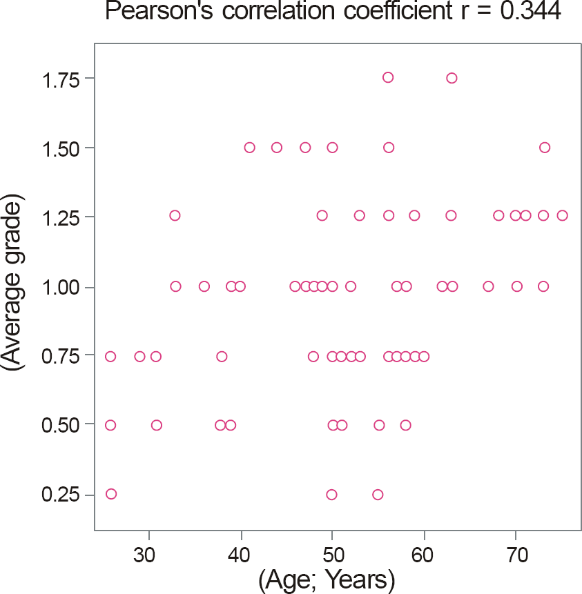 | Figure 2.The relationship between age and average grade of pingueculae ( p < 0.001). The average grade of pingueculae was defined as an average of each pinguecula that exists in both eyes. |
Table 1.
Grading system for pinguecula
Table 2.
Clinical profile of the pinguecula patient
Table 3.
Profile of pinguecula by location
| Number of patients (%) (n = 120) | |||
|---|---|---|---|
| Nasal and temporal of both eyes (4 quadrants) | 62 (52) | ||
| Nasal and temporal of an eye and nasal or temporal of an eye (3 quadrants) | 20 (17) | ||
| Nasal of both eyes (2 quadrants) | 20 (17) | ||
| Temporal of both eyes (2 quadrants) | 4 (3) | ||
| Nasal and temporal of an eye (2 quadrants) | 4 (3) | ||
| Nasal of an eye (1 quadrant) | 6 (5) | ||
| Temporal of an eye (1 quadrant) | 4 (3) | ||
| Nasal | Temporal | p-value | |
| Number of pinguecula of both eyes (%) (n = 374) | 208 (56) | 166 (44) | |
| Number of Pinguecula grade | |||
| P (1); mild or moderate pinguecula (%) | 132 (63.5) | 138 (83.1) | |
| P (2); severe pinguecula (%) | 76 (36.5) | 28 (16.9) | |
| Grade of pinguecula (mean ± SD) | 1.18 ± 0.648 | 0.81 ± 0.626 | <0.001* |
| Number of color | |||
| White (%) | 68 (32.7) | 82 (49.4) | |
| Yellowish white (%) | 92 (44.2) | 64 (38.6) | |
| Yellow (%) | 48 (23.1) | 20 (12) | |
| Brown (%) | 0 | 0 | |
| Number of elevated appearance | <0.001† | ||
| Flat (%) | 48 (23.1) | 94 (56.6) | |
| Slight elevation (%) | 94 (45.2) | 58 (34.9) | |
| Noticeable elevation (%) | 66 (31.7) | 14 (8.4) | |
| Number of vascularized appearance | |||
| Mild (0-5; %) | 154 (74) | 150 (90.4) | |
| Moderate (6-10; %) | 26 (12.5) | 12 (7.2) | |
| Severe (>11; %) | 28 (13.5) | 4 (2.4) | |
Table 4.
Profile of size by location
| Nasal | Temporal | p-value |
|---|---|---|
| Horizontal diameter (mm) 2.53 ± 1.25 | 2.76 ± 1.37 | 0.238* |
| (0.50-7.00) | (0.50-6.10) | |
| 2.40 ± 0.88 Vertical diameter (mm) | 2.14 ± 0.91 | 0.044* |
| (0.50-5.20) | (0.50-4.60) | |
| 2.47 ± 0.97 Average diameter (mm) | 2.45 ± 1.06 | 0.899* |
| (0.50-6.10) | (0.70-5.15) | |
| Horizontal diameter | Vertical diameter | p-value |
| Nasal and temporal of pinguecula (mm) 2.63 ± 1.30 | 2.28 ± 0.89 | 0.003* |
| (0.50-7.00) | (0.50-5.20) |
 XML Download