

 PDF
PDF ePub
ePub Citation
 Print
Print


Abstract
Purpose
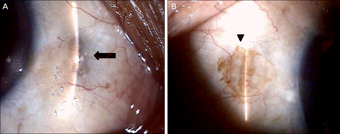
To report a case of bilateral scleral hyaline plaques in an elderly male patient. Scleral hyaline plaques are commonly noticed but rarely diagnosed. These are typical areas of hyalinization over the insertions of horizontal rectus muscle.
Case summary
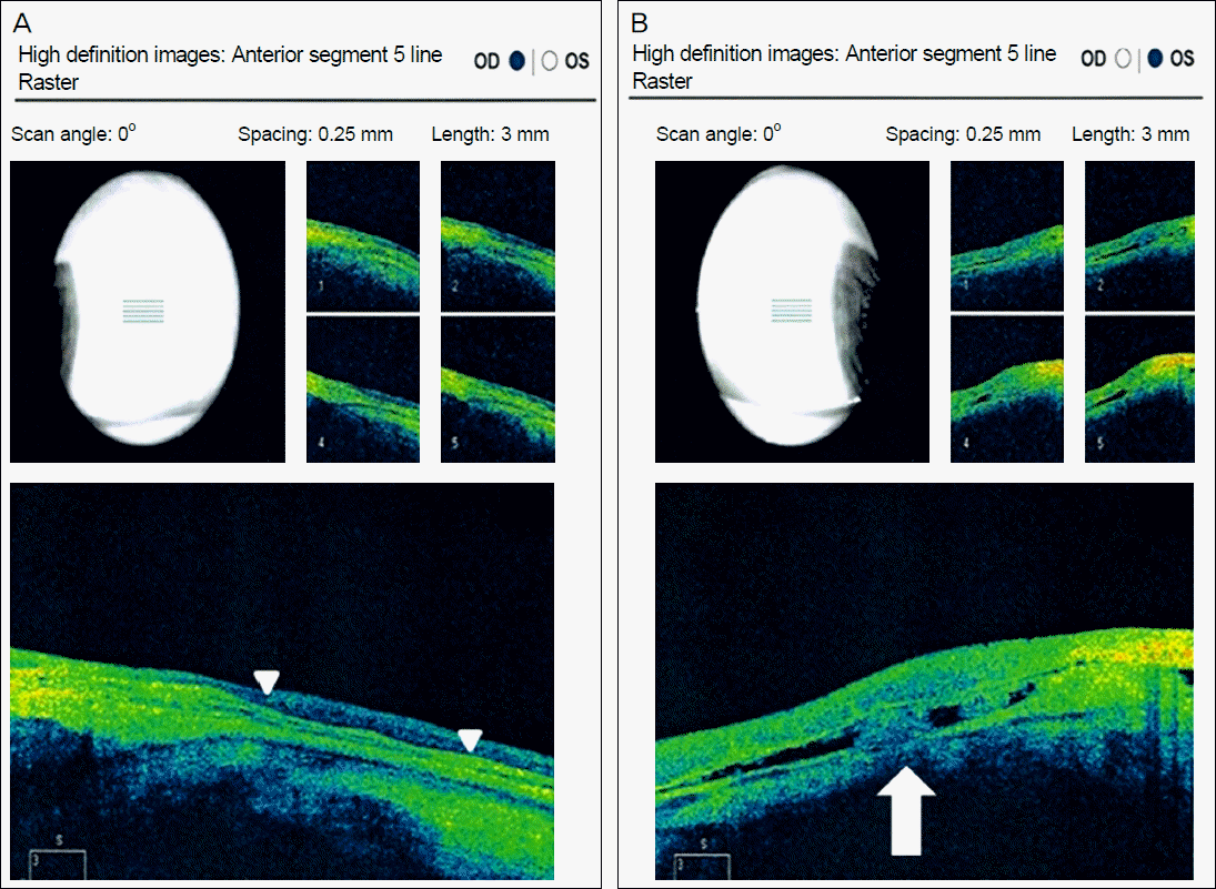
A 75-year-old man presented to the hospital complaining of scleral color change. External examination of both eyes revealed oval, slate-gray lesions measuring approximately 3.5 mm in diameter, located just anterior to the insertion of the medial rectus muscle in both eyes. On anterior segment optical coherence tomography of the lesions, thinned sclera and plaque were observed. There was no change in the lesion on the 3-year follow-up visit.
J Korean Ophthalmol Soc 2013;54(4):671-674
Go to : 
References
1. Moseley I. Spots before the eyes: a prevalence and clinicoradio-logical study of senile scleral plaques. Clin Radiol. 2000; 55:198–206.

2. Watson P. Diseases of the sclera and episclera. Tasman W, Jaeger EA, editors. Duane's Clinical Ophthalmology. Philadelphia: Lippincott-Raven;1997. 4:p. 40.
3. Donoso LA, Shields JA, Nagy RM. Epibulbar lesions simulating extraocular extension of uveal melanomas. Ann Ophthalmol. 1982; 14:1120–3.
4. Soong HK, McKenney MJ, Wolter JR. Adrenochrome staining of senile plaque resembling malignant melanoma. Am J Ophthalmol. 1986; 101:380.
5. Hillenkamp J, Sundmacher R, Sellmer R, Witschel H. Sequestrating senile scleral plaque simulating “necrotizing scleritis”Surgical management. Klin Monbl Augenheilkd. 2000; 216:177–80.
6. Foster CS, de la Maza MS. Noninflammatory diseases of the sclera. Foster CS, de la Maza MS. The Sclera. New York: Springer-Verlag;1994. p. 283.
7. Choi WS, Lee GJ, Park YJ, Lee KW. Scleral graft, free conjunctival autograft using tissue adhesive and temporary amniotic membrane transplantation in scleromalacia. J Korean Ophthalmol Soc. 2011; 52:1405–13.
8. Scroggs MW, Klintworth GK. Senile scleral plaques: a histopatho-logic study using energy-dispersive x-ray microanalysis. Hum Pathol. 1991; 22:557–62.
9. Murthy SI, Sangwan VS. Bilateral senile scleral plaques mimicking post-inflammatory scleral ectasia. Indian J Ophthalmol. 2004; 52:59–60.
10. Lyall DA, Srinivasan S. Scleral perforation secondary to a spontaneously dehisced senile scleral plaque: clinical features and management. Clin Experiment Ophthalmol. 2010; 38:533–4.
Go to :
 XML Download