

 PDF
PDF ePub
ePub Citation
 Print
Print


Abstract
Purpose
To report a case of Pseudomonas fluorescens infection following endoscopic dacryocystorhinostomy and silicone tube intubation in a healthy patient who was using steroid nasal spray. In addition, a literature review is conducted.
Case summary
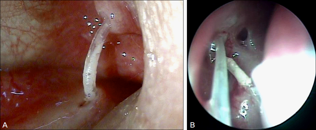
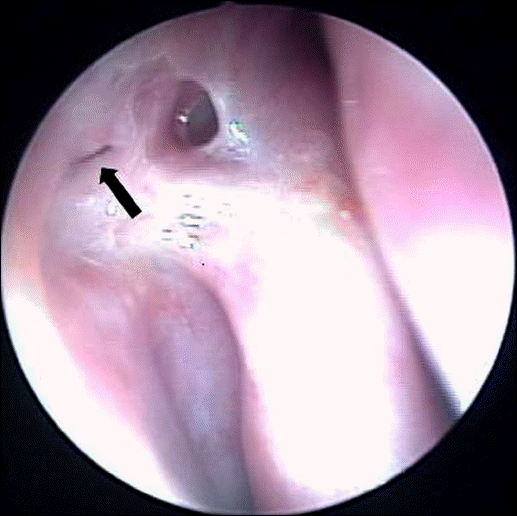
A 72-year-old female patient came to our clinic with tearing and hyperemia in the right eye. Ten months prior, she had undergone endoscopic dacryocystorhinostomy and silicone tube intubation due to nasolacrimal duct obstruction in the right eye. Six months after the first operation, dacryocystorhinostomy revision with silicone tube exchange was performed due to obstruction of the nasal bony orifice. In addition, the patient was using a steroid nasal spray. On slit lamp examination, conjunctival injection, marked inflammation and punctal edema around the tube were observed. The silicone tube was removed and the tube cultured. Pseudomonas fluorescens was isolated from the tube contents. The patients was treated with topical 0.3% gatifloxacin 4 times a day, methylol cephalexin lysinate 1000 mg 3 times a day and the nasal spray was discontinued. Two weeks later, all symptoms were resolved after treatment with antibiotic treatment.
References
1. Bartley GB. Acquired lacrimal drainage obstruction: an etiologic classification system, case reports, and a review of the literature. Part 3. Ophthal Plast Reconstr Surg. 1993; 9:11–26.
2. Tarbet KJ, Custer PL. External dacryocystorhinostomy. Surgical success, patient satisfaction, and economic cost. Ophthalmology. 1995; 102:1065–70.

3. Walland MJ, Rose GE. Factors affecting the success rate of open lacrimal surgery. Br J Ophthalmol. 1994; 78:888–91.
4. Becker BB. Dacryocystorhinostomy without flaps. Ophthalmic Surg. 1988; 19:419–27.
5. Ostler HB, Ostler MW. Diseases of the external eye and adnexa: a text and atlas. 1st ed.1. Baltimore: Williams & Wilkins;1993. p. 294–300.
6. Gershman MD, Kennedy DJ, Noble-Wang J, et al. Multistate outbreak of Pseudomonas fluorescens bloodstream infection after exposure to contaminated heparinized saline flush prepared by a compounding pharmacy. Clin Infect Dis. 2008; 47:1372–9.
7. Welham RA, Henderson PH. Results of dacryocystorhinostomy analysis of causes for failure. Trans Ophthalmol Soc U K. 1973; 93:601–9.
8. Gibbs DC. New probe for the intubation of lacrimal canaliculi with silicone rubber tubing. Br J Ophthalmol. 1967; 51:198.
9. Quickert MH, Dryden RM. Probes for intubation in lacrimal drainage. Trans Am Acad Ophthalmol Otolaryngol. 1970; 74:431–3.
10. Jordan DR, Nerad JA. An acute inflammatory reaction to silicone stents. Ophthal Plast Reconstr Surg. 1987; 3:147–50.
11. Carroll RP. Acute inflammatory reaction to silicone stents. Ophthal Plast Reconstr Surg. 1989; 5:71.
12. Jung BY, Kim YD. Canaliculitis after dacryocystorhinostomy with silicone tubes. J Korean Ophthalmol Soc. 2008; 49:390–5.
13. Ruby AJ, Lissner GS, O'Grady R. Surface reaction on silicone tubes used in the treatment of nasolacrimal drainage system obstruction. Ophthalmic Surg. 1991; 22:745–8.
14. Owji N, Khalili MR. Normalization of conjunctival flora after dacryocystorhinostomy. Ophthal Plast Reconstr Surg. 2009; 25:136–8.
15. Park HJ, Yi GY, Moon NJ. Bacteriologic study on normal conjunctival flora and change of antibiotic susceptability. J Korean Ophthalmol Soc. 2001; 42:817–24.
16. Wong V, Levi K, Baddal B, et al. Spread of Pseudomonas fluorescens due to contaminated drinking water in a bone marrow transplant unit. J Clin Microbiol. 2011; 49:2093–6.
 XML Download