

 PDF
PDF ePub
ePub Citation
 Print
Print


Abstract
Purpose
Eccrine ductal carcinoma is an extremely rare tumor that arises in the eccrine sweat glands. The authors of the present study describe a case of an eyelid mass diagnosed as eccrine ductal carcinoma.
Case summary
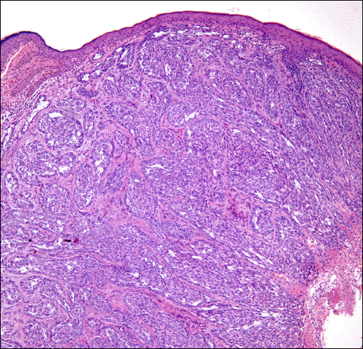
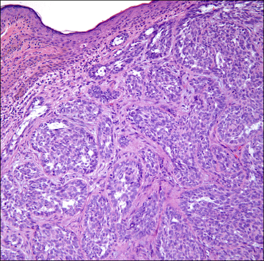
A 74-year-old woman visited our institute with a 3-month history of a mass in the left medial canthus. The lesion appeared as a solitary nodule with central ulceration. A magnetic resonance imaging (MRI) of the orbit showed a relatively well enhanced 0.8 cm × 0.8 cm-sized ovoid soft tissue mass. A mass excision was performed under frozen section control. The tumor was completely excised with margin clearance and medial canthal reconstruction was performed. Histopathological examination revealed a tumor composed of numerous duct-like structures lined with pleomorphic cuboidal epithelium that was diagnosed as eccrine ductal carcinoma of the eyelid.
Go to : 
References
1. Wick MR, Goellner JR. Wolfe JT 3rd, Su WP. Adnexal carcinomas of the skin. I. Eccrine carcinomas. Cancer. 1985; 56:1147–62.
2. Krishnakumar S, Mohan ER, Babu K, et al. Eccrine duct carcinoma of the eyelid mimicking meibomian carcinoma: Clinicopathological study of a case. Surv Ophthalmol. 2003; 48:439–46.

3. Urso C, Bondi R, Paglierani M, et al. Carcinomas of sweat glands: report of 60 cases. Arch Pathol Lab Med. 2001; 125:498–505.
4. Kacker A, Shaha AR. Ductal eccrine carcinoma arising in the post-aural area. Ear Nose Throat J. 1999; 78:576–7.
5. Shin J, Koh JK, Kim KH, et al. A clinicopathologic study on eccrine tumors. Korean J Dermatol. 2006; 44:1273–83.
6. Elder DE, Elenitsas R, Jaworsky C, Johnson Jr B. Lever's histo-pathology of the skin. 9th ed. Philadelphia: Lippincott Williams & Wilkins;2005. p. 899–914.
7. Ohnishi T, Kaneko S, Egi M, et al. Syringoid eccrine carcinoma: report of a case with immunohistochemical analysis of cytokeratin expression. Am J Dermatopathol. 2002; 24:409–13.
8. Weber PJ, Hevia O, Gretzula JC, Rabinovitz HC. Primary mucinous carcinoma. J Dermatol Surg Oncol. 1988; 14:170–2.
9. Herrero J, Monteagudo C, Jordá E, Llombart-Bosch A. Squamoid eccrine ductal carcinoma. Histopathology. 1998; 32:478–80.
10. Voutsadakis IA, Bruckner HW. Eccrine sweat gland carcinoma: a case report and review of diagnosis and treatment. Conn Med. 2000; 64:263–6.
11. Park BW, Kim SI, Lee KS, Yang WI. Ductal eccrine carcinoma presenting as a Paget's disease-like lesion of the breast. Breast J. 2001; 7:358–62.
12. Urso C, Paglierani M, Bondi R. Histologic spectrum of carcinomas with eccrine ductal differentiation (sweat-gland ductal carcinomas). Am J Dermatopathol. 1993; 15:435–40.
13. Swanson PE, Cherwitz DL, Neumann MP, Wick MR. Eccrine sweat gland carcinoma: an histologic and immunohistochemical study of 32 cases. J Cutan Pathol. 1987; 14:65–86.
14. Wick MR, Ockner DM, Mills SE, et al. Homologous carcinomas of the breasts, skin, and salivary glands. A histologic and immunohistochemical comparison of ductal mammary carcinoma, ductal sweat gland carcinoma, and salivary duct carcinoma. Am J Clin Pathol. 1998; 109:75–84.
15. Swanson PE, Mazoujian G, Mills SE, et al. Immunoreactivity for estrogen receptor protein in sweat gland tumors. Am J Surg Pathol. 1991; 15:835–41.
16. Holds JB, Haines JH, Mamalis N, et al. Mucinous adenocarcinoma of the orbit arising from a stable, benign-appearing eyelid nodule. Ophthalmic Surg. 1990; 21:163–6.
17. Cruz DJ. Sweat gland carcinomas: a comprehensive review. Semin Diagn Pathol. 1987; 4:38–74.
Go to :
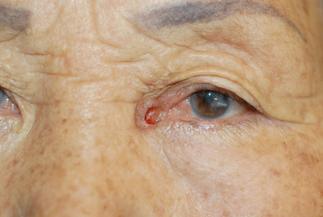 | Figure 1.Clinical photograph showing a firm mass with central ulceration in the medial canthal area of the left eye. |
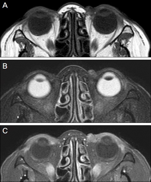 | Figure 2.(A) T1-weighted axial image demonstrating a poorly marginated mass with low signal intensity. (B) T2-weighted axial image demonstrating a heterogeneous high signal intensity in the left medial canthal area. (C) Contrast enhanced T1-weighted images demonstrating a relatively well enhancing soft tissue mass in the left medial canthal area. |
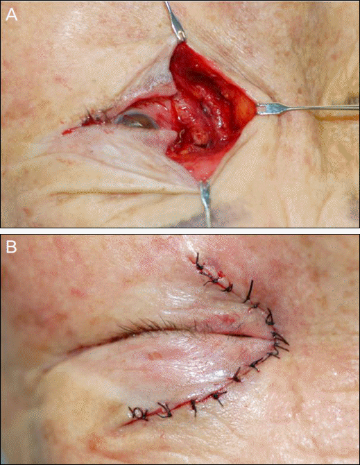 | Figure 3.(A) Intraoperative photography showing a widely excised lesion with large defect. (B) A medial canthal reconstruction using advancement flap. |
 XML Download