Abstract
Purpose
To evaluate the stability and optical performance of the newly developed single-piece aspheric intraocular lens (IOL) by comparing the clinical outcome of the aspheric IOL with the new optic profile design (HOYA iSert, HOYA iMics) and the aspheric IOL (Akreos MI60), which has been proven effective and safe.
Methods
iSert, iMics, and MI60 were inserted into 55 eyes, 60 eyes, and 50 eyes, respectively, after microincision phacoemulsification cataract surgery. Best corrected visual acuity (BCVA), refraction in spherical equivalent, anterior chamber depth (ACD), total higher order aberration (HOA), contrast sensitivity, and surgically induced astigmatism (SIA) were measured and each IOL was evaluated on the functional stability, anterior-posterior stability, centration in the capsular bag, and quality of vision.
Results
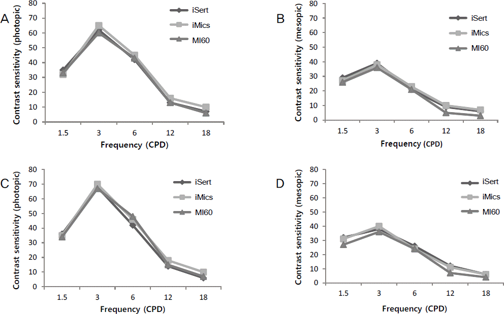
No statistical differences in preoperative and postoperative BCVA among the 3 IOL groups were observed, however, MI60 showed significant myopic shift postoperatively. Anterior-posterior stability assessed with postoperative change in refractive error and ACD was slightly lower in the MI60 group. In terms of vision quality, while total aberration, total HOA, coma aberration, and contrast sensitivity for the 3 IOLs were not different significantly, spherical aberration of the MI60 group was higher than the other groups at 6 months postoperative. SIA was significantly increased in eyes implanted with iSert than in eyes with iMics or MI60 at 1 month postoperatively, however, the differences were no longer evident after 3 months postoperatively.
References
1. Alió J, Rodríguez-Prats JL, Galal A, Ramzy M. Outcomes of microincision cataract surgery versus coaxial phacoemulsification. Ophthalmology. 2005; 112:1997–2003.

2. Kurz S, Krummenauer F, Gabriel P, et al. Biaxial microincision versus coaxial small-incision clear cornea cataract surgery. Ophthalmology. 2006; 113:1818–26.

3. Shin CJ, Lee JE, Lee JH, et al. Clinical outcomes after micro-incision cataract surgery and in-the-bag implantation of a new intraocular lens. J Korean Ophthalmol Soc. 2010; 51:677–83.

4. Chantra S, Pachimkul P, Naripthaphan P. Wavefront and ocular spherical aberration after implantation of different types of aspheric intraocular lenses based on corneal spherical aberration. J Med Assoc Thai. 2011; 94(Suppl 2):S71–5.
5. Holladay JT, Piers PA, Koranyi G, et al. A new intraocular lens design to reduce spherical aberration of pseudophakic eyes. J Refract Surg. 2002; 18:683–91.

6. Nochez Y, Favard A, Majzoub S, Pisella PJ. Measurement of cor-neal aberrations for customisation of intraocular lens asphericity: impact on quality of vision after micro-incision cataract surgery. Br J Ophthalmol. 2010; 94:440–4.

7. Kohnen T, Klaproth OK, Bühren J. Effect of intraocular lens asphericity on quality of vision after cataract removal: an intra-individual comparison. Ophthalmology. 2009; 116:1697–706.
8. Nejima R, Miyai T, Kataoka Y, et al. Prospective intrapatient comparison of 6.0-millimeter optic single-piece and 3-piece hydro-phobic acrylic foldable intraocular lenses. Ophthalmology. 2006; 113:585–90.

9. Caporossi A, Casprini F, Tosi GM, Baiocchi S. Preliminary results of cataract extraction with implantation of a single-piece AcrySof intraocular lens. J Cataract Refract Surg. 2002; 28:652–5.

10. Eppig T, Scholz K, Löffler A, et al. Effect of decentration and tilt on the image quality of aspheric intraocular lens designs in a model eye. J Cataract Refract Surg. 2009; 35:1091–100.

11. Mester U, Sauer T, Kaymak H. Decentration and tilt of a single-piece aspheric intraocular lens compared with the lens position in young phakic eyes. J Cataract Refract Surg. 2009; 35:485–90.

12. Casprini F, Balestrazzi A, Tosi GM, et al. Glare disability and spherical aberration with five foldable intraocular lenses: a prospective randomized study. Acta Ophthalmol Scand. 2005; 83:20–5.

13. Yoon JU, Chung JL, Hong JP, et al. Comparison of wavefront analysis and visual function between monofocal and multifocal aspheric intraocular lenses. J Korean Ophthalmol Soc. 2009; 50:195–201.

14. Kang IS YI, You IC, Park YG, Yoon KC. Comparison of visual function among aspheric intraocular lenses. J Korean Ophthalmol Soc. 2009; 50:691–7.

15. Lee JY, Lee SH, Chung SK. Decentration, tilt and anterior chamber depth: aspheric vs spheric acrylic intraocular lens. J Korean Ophthalmol Soc. 2009; 50:852–7.

16. Altmann GE, Nichamin LD, Lane SS, Pepose JS. Optical performance of 3 intraocular lens designs in the presence of decentration. J Cataract Refract Surg. 2005; 31:574–85.

17. Matsushima H. Characteristics of iMics1. Japanese J Ophthalmic Surg. 2009; 22:1–7.
18. Wirtitsch MG, Findl O, Menapace R, et al. Effect of haptic design on change in axial lens position after cataract surgery. J Cataract Refract Surg. 2004; 30:45–51.

19. Petternel V, Menapace R, Findl O, et al. Effect of optic edge design and haptic angulation on postoperative intraocular lens position change. J Cataract Refract Surg. 2004; 30:52–7.

20. Hayashi K, Yoshida M, Hayashi H. Postoperative corneal shape changes: microincision versus small-incision coaxial cataract surgery. J Cataract Refract Surg. 2009; 35:233–9.

21. Tong N, He JC, Lu F, et al. Changes in corneal wavefront aberrations in microincision and small-incision cataract surgery. J Cataract Refract Surg. 2008; 34:2085–90.

22. Hayashi K, Hayashi H. Visual function in patients with yellow tinted intraocular lenses compared with vision in patients with non-tinted intraocular lenses. Br J Ophthalmol. 2006; 90:1019–23.

23. Neumaier-Ammerer B, Felke S, Hagen S, et al. Comparison of visual performance with blue light-filtering and ultraviolet light-filtering intraocular lenses. J Cataract Refract Surg. 2010; 36:2073–9.

24. Henderson BA, Grimes KJ. Blue-blocking IOLs: a complete review of the literature. Surv Ophthalmol. 2010; 55:284–9.

25. Wang H, Wang J, Fan W, Wang W. Comparison of photochromic, yellow, and clear intraocular lenses in human eyes under photopic and mesopic lighting conditions. J Cataract Refract Surg. 2010; 36:2080–6.

26. Yamaguchi T, Negishi K, Ono T, et al. Feasibility of spherical aberration correction with aspheric intraocular lenses in cataract surgery based on individual pupil diameter. J Cataract Refract Surg. 2009; 35:1725–33.

27. Yamaguchi T, Negishi K, Ohnuma K, Tsubota K. Correlation between contrast sensitivity and higher-order aberration based on pupil diameter after cataract surgery. Clin Ophthalmol. 2011; 5:1701–7.

28. Mester U, Kaymak H. [The aspheric blue light filter IOL AcrySof IQ compared to the AcrySof SA60AT : influence of IOL power, pupil diameter, and corneal asphericity on postoperative spherical.
29. Solomon JD. Outcomes of corneal spherical aberration-guided cataract surgery measured by the OPD-scan. J Refract Surg. 2010; 26:863–9.

30. Negishi K, Kodama C, Yamaguchi T, et al. Predictability of ocular spherical aberration after cataract surgery determined using pre-operative corneal spherical aberration. J Cataract Refract Surg. 2010; 36:756–61.

31. Mutlu FM, Bilge AH, Altinsoy HI, Yumusak E. The role of capsulotomy and intraocular lens type on tilt and decentration of polymethylmethacrylate and foldable acrylic lenses. Ophthalmologica. 1998; 212:359–63.

32. Hayashi K, Harada M, Hayashi H, et al. Decentration and tilt of polymethyl methacrylate, silicone, and acrylic soft intraocular lenses. Ophthalmology. 1997; 104:793–8.

33. Nanavaty MA, Spalton DJ, Boyce J, et al. Edge profile of commercially available square-edged intraocular lenses. J Cataract Refract Surg. 2008; 34:677–86.

34. Werner L, Müller M, Tetz M. Evaluating and defining the sharpness of intraocular lenses: microedge structure of commercially available square-edged hydrophobic lenses. J Cataract Refract Surg. 2008; 34:310–7.
Figure 1.
Comparison of postoperative contrast sensitivity in photophic and mesopic conditions (M ean). The asterisks (*) indicate the differences in values were significant (p < 0.05) among three groups. (A) Contrast sensitivity in photopic condition at postoperative 1 month. (B) Contrast sensitivity in mesopic condition at postoperative 1 month. (C) Contrast sensitivity in photopic condition at postoperative 3 months. (D) Contrast sensitivity in mesopic condition at postoperative 3 months.

Table 1.
Comparison of preoperative characteristics among three groups
| HOYA iSert PC-60AD (n = 55) | HOYA iMics NY-60 (n = 60) | Akreos MI60 (n = 50) | p-value | |
|---|---|---|---|---|
| Age (years) | 70.2 ± 8.6 | 68.6 ± 12.3 | 66.7 ± 10.5 | 0.231 |
| Sex (Male/Female) | 30/25 | 29/31 | 28/22 | 0.155* |
| BCVA (log MAR) | 0.50 ± 0.24 | 0.45 ± 0.30 | 0.46 ± 0.44 | 0.126 |
| Manifest refraction (Diopter) | -0.88 ± 2.95 | -1.01 ± 2.84 | -0.83 ± 2.72 | 0.305 |
| Axial Length (mm) | 23.2 ± 0.8 | 23.4 ± 0.9 | 23.6 ± 1.3 | 0.255 |
| IOL power (Diopters) | 20.5 ± 2.6 | 21.1 ± 2.3 | 20.9 ± 2.2 | 0.520 |
| Target refraction (Diopter) | -0.20 ± 0.51 | -0.25 ± 0.79 | -0.22 ± 0.63 | 0.385 |
| ACD (mm) | 2.60 ± 0.51 | 2.72 ± 0.36 | 2.76 ± 0.43 | 0.177 |
| Pupil diameter (mm) under mesopic condition | 4.64 ± 0.66 | 4.52 ± 0.60 | 4.62 ± 0.55 | 0.441 |
Table 2.
Comparison of postoperative total aberration, total high order aberration, spherical aberration, and coma aberration for three different IOLs by 6 months postoperative


 PDF
PDF ePub
ePub Citation
 Print
Print


 XML Download