

 PDF
PDF ePub
ePub Citation
 Print
Print


Abstract
Purpose
The present study investigates the effects of pharmacologic pupil dilatation on ocular, corneal and internal aberrations.
Methods
Sixty-two right eyes of 62 healthy participants were included in the present study. Ocular, corneal and internal aberrations were measured with a KR-1W wavefront aberrometer (Topcon Corp., Tokyo, Japan) before mydriasis in mesopic conditions. After pupil dilatation with a mydriatic drug (phenylephrine chloride 0.5% + tropicamide 0.5%) (Mydrin-P, Santen, Osaka, Japan), the measurements were repeated. The wavefront data of 4-mm and 6-mm diameter zones were analyzed. The changes of aberrations before and after mydriasis were evaluated by paired t-test.
Results
The values of ocular, corneal and internal spherical aberrations before and after mydriasis on the 4-mm diameter pupil zone were not statistically significantly different. On the 6-mm diameter zone, the ocular and internal spherical aberrations were statistically significantly different (p = 0.025, p = 0.002, respectively, paired t-test). However, the corneal aberrations did not show significant changes. The internal aberrations average before mydriasis was -0.043 (±0.21) μm and was shifted in a negative direction to -0.093 (±0.17) μm after mydriasis. The ocular aberrations average also changed toward negative after mydriasis. The high-order aberrations and astigmatism did not change significantly.
References
1. Cashell GT. A short history of spectacles. Proc R Soc Med. 1971; 64:1063–4.

2. Born M, Wolf E. Principles of optics, 7th ed. Cambridge: Cambridge University Press;1999. p. 523–5.
3. Molebny VV, Pallikaris IG, Naoumidis LP, et al. Retina ray-tracing technique for eye-refraction mapping. Proc SPIE. 1997; 2971:175–83.
4. Molebny VV, Panagopoulou SI, Molebny SV, et al. Principles of ray tracing aberrometry. J Refract Surg. 2000; 16:S572–5.
5. MacRae S, Fujieda M. Slit skiascopic-guided ablation using the Nidek laser. J Refract Surg. 2000; 16:S576–80.
6. Mrochen M, Kaemmerer M, Mierdel P, et al. Principles of Tscherning aberrometry. J Refract Surg. 2000; 16:S570–1.
7. Moreno-Barriuso E, Navarro R. Laser Ray Tracing versus Hartmann-Shack sensor for measuring optical aberrations in the human eye. J Opt Soc Am A Opt Image Sci Vis. 2000; 17:974–85.
8. Thibos LN. Principles of Hartmann-Shack aberrometry. J Refract Surg. 2000; 16:S563–5.
9. Liang J, Grimm B, Goelz S, Bille JF. Objective measurement of wave aberrations of the human eye with the use of a Hartmann-Shack wave-front sensor. J Opt Soc Am A Opt Image Sci Vis. 1994; 11:1949–57.
10. Ortiz D, Alió JL, Bernabéu G, Pongo V. Optical performance of monofocal and multifocal intraocular lenses in the human eye. J Cataract Refract Surg. 2008; 34:755–62.
11. Giessler S, Hammer T, Duncker GI. [Aberrometry due dilated pupilsWhich mydriatic should be used?]. Klin Monbl Augenheilkd. 2002; 219:655–9.
12. Carkeet A, Velaedan S, Tan YK, et al. Higher order ocular aberrations after cycloplegic and non-cycloplegic pupil dilation. J Refract Surg. 2003; 19:316–22.
13. Taneri S, Oehler S, Azar DT. Influence of mydriatic eye drops on wavefront sensing with the Zywave aberrometer. J Refract Surg. 2011; 27:678–85.
14. Ahn SM, Seok SS, Park CY. Considering spherical aberration in choosing the wavefront map for laser vision correction. J Korean Ophthalmol Soc. 2011; 52:147–56.
15. Piñero DP, Juan JT, Alió JL. Intrasubject repeatability of internal aberrometry obtained with a new integrated aberrometer. J Refract Surg. 2011; 27:509–17.
16. Brown N. The change in shape and internal form of the lens of the eye on accommodation. Exp Eye Res. 1973; 15:441–59.
17. Dubbelman M, Van der Heijde GL, Weeber HA. Change in shape of the aging human crystalline lens with accommodation. Vision Res. 2005; 45:117–32.
18. Dubbelman M, Van der Heijde GL, Weeber HA, Vrensen GF. Changes in the internal structure of the human crystalline lens with age and accommodation. Vision Res. 2003; 43:2363–75.
Figure 1.
Comparison of spherical aberrations (SA) before and after mydriasis (in micrometer). The ocular and internal spherical aberrations were statistically significantly different on 6-mm diameter zone (p = 0.025, p = 0.002, respectively, paired t-test). Before = before mydriasis, at mesopic conditions, 8 lux; After = after pharmacologically induced mydriasis.
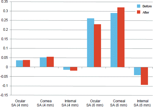
Figure 2.
Comparison of high order aberrations (HOA) before and after mydriasis (in average root mean square; RMS). The high order aberrations did not change significantly after mydriasis. Before = before mydriasis, at mesopic conditions, 8 lux; After = after pharmacologically induced mydriasis.
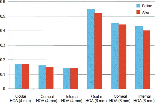
Figure 3.
Comparison of astigmatism (Astig.) before and after mydriasis (in diopter). The astigmatism did not change significantly after mydriasis. Before = before mydriasis, at mesopic conditions, 8 lux; After = after pharmacologically induced mydriasis.
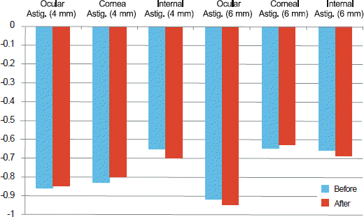
Table 1.
The average of ocular, corneal and internal spherical aberrations on 4-mm and 6-mm diameter zone before and after mydriasis. The ocular and internal spherical aberrations on 6-mm diameter zone were statistically significantly shifted in a negative direction after mydriasis
| Pupil size | Before* (μm) | After† (μm) | p-value | |
|---|---|---|---|---|
| 4-mm | Ocular SA | 0.037 ± 0.035 | 0.038 ± 0.039 | 0.857 |
| Corneal SA | 0.052 ± 0.029 | 0.056 ± 0.029 | 0.343 | |
| Internal SA | -0.014 ± 0.037 | -0.019 ± 0.032 | 0.228 | |
| 6-mm | Ocular SA | 0.26 ± 0.25 | 0.23 ± 0.25 | 0.025 |
| Corneal SA | 0.29 ± 0.21 | 0.32 ± 0.18 | 0.088 | |
| Internal SA | -0.043 ± 0.21 | -0.093 ± 0.17 | 0.002 |
Table 2.
The average of ocular, corneal and internal high order aberrations on 4-mm and 6-mm diameter zone before and after mydriasis. The high order aberrations were not statistically significantly different after mydriasis
| Pupil size | Before* (RMS‡) | After† (RMS) | p-value | |
|---|---|---|---|---|
| 4-mm | Ocular HOA | 0.16 ± 0.084 | 0.16 ± 0.089 | 0.906 |
| Corneal HOA | 0.16 ± 0.071 | 0.15 ± 0.061 | 0.241 | |
| Internal HOA | 0.13 ± 0.098 | 0.13 ± 0.097 | 0.945 | |
| 6-mm | Ocular HOA | 0.54 ± 0.33 | 0.52 ± 0.33 | 0.139 |
| Corneal HOA | 0.48 ± 0.26 | 0.48 ± 0.26 | 0.921 | |
| Internal HOA | 0.42 ± 0.24 | 0.39 ± 0.21 | 0.267 |
Table 3.
The average of ocular, corneal and internal astigmatism on 4-mm and 6-mm diameter zone before and after mydriasis. The astigmatism was not statistically significantly different after mydriasis
| Pupil size | Before* (D) | After† (D) | p-value | |
|---|---|---|---|---|
| 4-mm | Ocular Astig. | -0.86 ± 0.49 | -0.85 ± 0.46 | 0.936 |
| Corneal Astig. | -0.83 ± 0.54 | -0.80 ± 0.54 | 0.409 | |
| Internal Astig. | -0.65 ± 0.37 | -0.70 ± 0.39 | 0.229 | |
| 6-mm | Ocular Astig. | -0.92 ± 0.49 | -0.95 ± 0.55 | 0.417 |
| Corneal Astig. | -0.65 ± 0.43 | -0.63 ± 0.47 | 0.579 | |
| Internal Astig. | -0.66 ± 0.39 | -0.69 ± 0.43 | 0.427 |
 XML Download