

 PDF
PDF ePub
ePub Citation
 Print
Print


Abstract
Purpose
To investigate the effects of Nasopore as a nasal packing material on the surgical success rate and prevalence of postoperative complications after endonasal dacryocystorhinostomy (DCR).
Methods
The present study included a total of 558 patients (699 eyes) with primary acquired nasolacrimal duct obstruction who underwent endonasal DCR; 227 eyes were packed with Nasopore and 472 eyes were packed with Merocel. The surgical success rate and postoperative complications such as synechiae, granulation, wound healing (osteal mucosal epithelium epithelialization), postoperative bleeding, infection, and revision rate were compared between the packing materials. Results: The surgical success rate of the Nasopore group (99.1%, 98.6%) showed significantly better results than the Merocel group (97.2%, 95.1%) at postoperative 1 and 3 months (p = 0.04, 0.03 Pearson chi-square test), whereas there was no statistically significant difference between the 2 groups in postoperative surgical success rate at 1 week and 6 months. In comparison of postoperative complications, the Nasopore group (0%) showed a lower incidence of delayed wound healing (delayed epithelialization of osteal mucosal epithelium) than the Merocel group (2.3%; p = 0.013), whereas there was no difference in granulation, synechiae, postoperative bleeding, infection and revision rate (p > 0.05).
Go to : 
References
1. Weber R, Keerl R, Hochapfel F, et al. Packing in endonasal surgery. Am J Otolaryngol. 2001; 22:306–20.

2. Fairbanks DN. Complications of nasal packing. Otolaryngol Head Neck Surg. 1986; 94:412–5.
3. von Schoenberg M, Robinson P, Ryan R.Nasal packing after routine nasal surgery--is it justified? J Laryngol Otol. 1993; 107:902–5.
4. Weber R, Hochapfel F, Draf W. Packing and stents in endonasal surgery. Rhinology. 2000; 38:49–62.
5. Anderson RL. Gelfoam packing after dacryocystorhinostomy. Arch Ophthalmol. 1977; 95:520.
6. Basmak H, Cakli H, Sahin A, et al. Comparison of endocanalicular laser dacryocystorhinostomy with and without endonasal procedures. Graefes Arch Clin Exp Ophthalmol. 2011; 249:737–43.
Go to :
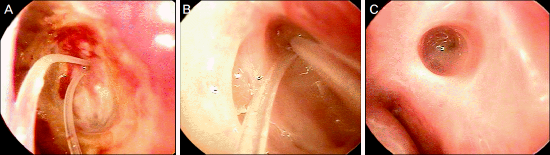 | Figure 1.Representative photographs showing (A) delayed epithelialization of osteal mucosal epithelium at 2 months, (B) normally recovered epithelialization of osteal mucocal epithelium at 2 months, (C) fully recovered mucosal epithelialization at 6 months. |
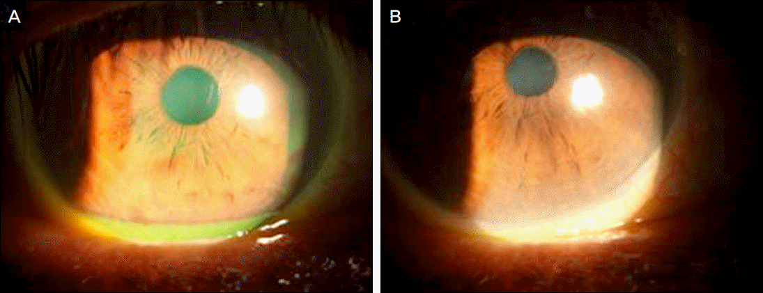 | Figure 2.Representative photographs showing tear meniscus height measured by slit-lamp examination and fluorescein staining. (A) Preoperative tear meniscus height, determined as 3 mm. (B) Tear meniscus height after tube removal, determined as 0.5 mm. |
Table 1.
Patient demographics & characteristics at baseline
| Group 1 (Merocel®) n = 472 | Group 2 (Nasopore®) n = 227 | p-value | |
|---|---|---|---|
| Age (years) | 56.77 ± 10.1 | 55.04 ± 11.8 | 0.06* |
| Gender | 0.06† | ||
| Male | 98 (20.8%) | 29 (12.8%) | |
| Female | 374 (79.2%) | 198 (87.2%) | |
| Side | 0.36† | ||
| OD | 235 (49.8%) | 109 (48.0%) | |
| OS | 237 (50.2%) | 118 (52.0%) | |
| Previous dacryocystitis history | 14 (3.0%) | 10 (4.4%) | 0.222† |
| Mean time to tube removal (week) | 16.41 ± 4.68 | 15.35 ± 3.42 | 0.002* |
Table 2.
Comparision of postoperative general complications
| Group 1 (Merocel®) n = 472 | Group 2 (Nasopore®) n = 227 | p-value | |
|---|---|---|---|
| Delayed wound healing (Delayed epithelialization) | 11 (2.3%) | 0 (0%) | 0.013* |
| Granuloma formation | 146 (30.9%) | 67 (30.3%) | 0.64* |
| Synechiae | 8 (1.7%) | 5 (2.2%) | 0.42* |
| Postoperative bleeding | 4 (0.8%) | 1 (0.4%) | 0.48* |
| Postoperative infection | 9 (1.9%) | 6 (2.6%) | 0.35* |
| Revision | 9 (1.9%) | 1 (0.4%) | 0.11* |
Table 3.
Comparison of surgical success rate
| Group 1 (Merocel®) n = 472 | Group 2 (Nasopore®) n = 227 | p-value | |
|---|---|---|---|
| Postoperative 1 week | 465/472 (98.5 %) | 223/227 (98.2%) | 0.87* |
| Postoperative 1 month | 459/472 (97.2%) | 225/227 (99.1%) | 0.04* |
| Postoperative 3 months | 443/466 (95.1%) | 204/207 (98.6%) | 0.03* |
| Postoperative 6 months | 167/182 (91.8%) | 34/41 (82.9%) | 0.09* |
 XML Download