Abstract
Purpose
To compare the outcomes of autogenous dermis fat grafting with different donor sites in the treatment of exposed porous orbital implants.
Methods
The present study retrospectively evaluated the medical records of 17 patients (17 anophthalmic eyes) who had undergone autogenous dermis fat grafting based on the diagnosis of exposed porous orbital implants and were regularly followed up for at least 12 months since the surgery from January 2001 to December 2010. The patients were divided into 2 groups (thigh and abdomen) according to the site of the donor grafting. The treatment outcome and complications were compared between the 2 groups.
Results
The success rate of thigh dermis fat grafting was 88.9% (8/9) and 100.0% (8/8) in the abdominal dermis fat grafting, and there was no statistically significant difference between the 2 groups (p = 1.000). Regarding ocular complications, graft tissue infection (thigh 11.1%, abdomen 0%) and superior sulcus deformity (thigh 22.2%, abdomen 25.0%) were present. Regarding donor site complications, tenderness (thigh 55.6%, abdomen 25.0%), dehiscence (thigh 22.2%, abdomen 25.0%) and scar formation (thigh 33.3%, abdomen 25.0%) were observed. In the gait associated complications, pain (thigh 55.6%, abdomen 25.0%) and limping (thigh 22.2%, abdomen 12.5%) were observed. The rate of all complications showed no statistically significant difference between the thigh dermis fat grafting and the abdominal dermis fat grafting (all p > 0.05).
Go to : 
References
1. Su GW, Yen MT. Current trends in managing the anophthalmic socket after primary enucleation and evisceration. Ophthal Plast Reconstr Surg. 2004; 20:274–80.

2. Viswanathan P, Sagoo MS, Olver JM. UK national survey of enucleation, evisceration and orbital implant trends. Br J Ophthalmol. 2007; 91:616–9.

4. Li T, Shen J, Duffy MT. Exposure rates of wrapped and unwrapped orbital implants following enucleation. Ophthal Plast Reconstr Surg. 2001; 17:431–5.

5. McNab A. Hydroxyapatite orbital implants. Experience with 100 cases. Aust N Z J Ophthalmol. 1995; 23:117–23.
6. Oestreicher JH, Liu E, Berkowitz M. Complications of hydroxyapatite orbital implants. A review of 100 consecutive cases and a comparison of Dexon mesh (polyglycolic acid) with scleral wrapping. Ophthalmology. 1997; 104:324–9.

7. Custer PL, Trinkaus KM. Porous implant exposure: Incidence, management, and morbidity. Ophthal Plast Reconstr Surg. 2007; 23:1–7.

8. Nunery WR, Heinz GW, Bonnin JM, et al. Exposure rate of hydroxyapatite spheres in the anophthalmic socket: histopathologic correlation and comparison with silicone sphere implants. Ophthal Plast Reconstr Surg. 1993; 9:96–104.
9. Remulla HD, Rubin PA, Shore JW, et al. Complications of porous spherical orbital implants. Ophthalmology. 1995; 102:586–93.

10. Yoon JS, Lew H, Kim SJ, Lee SY. Exposure rate of hydroxyapatite orbital implants a 15-year experience of 802 cases. Ophthalmology. 2008; 115:566–72.
11. Park MS, Kim KS, Baek SH, Lee TS. Management of exposed porous orbital implant with autogenous dermis graft. J Korean Ophthalmol Soc. 2001; 42:1127–32.
12. Hwang K, Kim DJ, Lee IJ. An anatomic comparison of the skin of five donor sites for dermal fat graft. Ann Plast Surg. 2001; 46:327–31.

13. Lee MJ, Khwarg SI, Choung HK, et al. Dermis-fat graft for treatment of exposed porous polyethylene implants in pediatric post-enucleation retinoblastoma patients. Am J Ophthalmol. 2011; 152:244–50.

14. Rosen HM, McFarland MM. The biologic behavior of hydroxyapatite implanted into the maxillofacial skeleton. Plast Reconstr Surg. 1990; 85:718–23.

15. Goldberg RA, Holds JB, Ebrahimpour J. Exposed hydroxyapatite orbital implants. Report of six cases. Ophthalmology. 1992; 99:831–6.

16. Kim YD, Goldberg RA, Shorr N, Steinsapir KD. Management of exposed hydroxyapatite orbital implants. Ophthalmology. 1994; 101:1709–15.

17. Buettner H, Bartley GB. Tissue breakdown and exposure associated with orbital hydroxyapatite implants. Am J Ophthalmol. 1992; 113:669–73.

18. Martin P, Ghabrial R. Repair of exposed hydroxyapatite orbital implant by a tarsoconjunctival pedicle flap. Ophthalmology. 1998; 105:1694–7.

19. Massry GG, Holds JB. Frontal periosteum as an exposed orbital implant cover. Ophthal Plast Reconstr Surg. 1999; 15:79–82.

20. Pelletier CR, Jordan DR, Gilberg SM. Use of temporalis fascia for exposed hydroxyapatite orbital implants. Ophthal Plast Reconstr Surg. 1998; 14:198–203.

21. Rosen CE. The Müller muscle flap for repair of an exposed hydroxyapatite orbital implant. Ophthal Plast Reconstr Surg. 1998; 14:204–7.

22. Soparkar CN, Patrinely JR. Tarsal patch-flap for orbital implant exposure. Ophthal Plast Reconstr Surg. 1998; 14:391–7.

23. Smith B, Petrelli R. Dermis-fat graft as a movable implant within the muscle cone. Am J Ophthalmol. 1978; 85:62–6.

24. Davis RE, Guida RA, Cook TA. Autologous free dermal fat graft. Reconstruction of facial contour defects. Arch Otolaryngol Head Neck Surg. 1995; 121:95–100.
25. van Gemert JV, Leone CR Jr.Correction of a deep superior sulcus with dermis-fat implantation. Arch Ophthalmol. 1986; 104:604–7.

26. Conley JJ, Clairmont AA. Dermal-fat-fascia grafts. Otolaryngology. 1978; 86((4 Pt 1)):ORL-641-9.

27. Nosan DK, Ochi JW, Davidson TM. Preservation of facial contour during parotidectomy. Otolaryngol Head Neck Surg. 1991; 104:293–8.

28. Leaf N, Zarem HA. Correction of contour defects of the face with dermal and dermal-fat grafts. Arch Surg. 1972; 105:715–9.

29. Grillner S, Nilsson J, Thorstensson A. Intra-abdominal pressure changes during natural movements in man. Acta Physiol Scand. 1978; 103:275–83.

Go to : 
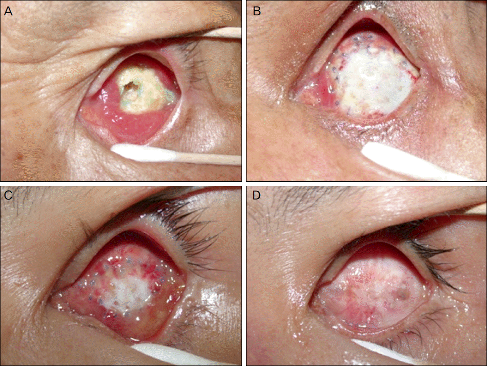
 | Figure 1.(A) Preoperative photograph of an exposed porous orbital implant at a 54-year-old male. (B) Post-operative (1 week after dermis fat graft) photograph. (C) Post-operative (1 month after dermis fat graft) photograph. (D) At post-operative 6 months after dermis fat graft, no evidence of re-exposure is observed. |
Table 1.
Clinical data of 17 patients with exposure of implant
| Case | Age/Sex | Diagnosis | Surgery | Implant | Exposure size | Donor site | Healing period (week) | F/U period (month) | Result |
|---|---|---|---|---|---|---|---|---|---|
| 1 | 20/M | Eyeball rupture | Enucleation | HAP | Large* | Thigh | 6 | 18 | No exposure |
| 2 | 70/M | Phthisis bulbi | Evisceration | HAP | Large | Thigh | 8 | 27 | No exposure |
| 3 | 50/M | Phthisis bulbi | Evisceration | HAP | Large | Thigh | 8 | 12 | No exposure |
| 4 | 42/M | Eyeball rupture | Enucleation | HAP | Large | Thigh | 6 | 13 | No exposure |
| 5 | 30/F | Eyeball rupture | Enucleation | HAP | Large | Thigh | 5 | 14 | No exposure |
| 6 | 58/F | Phthisis bulbi | Evisceration | Medpor® | Large | Thigh | 8 | 21 | No exposure |
| 7 | 60/M | Phthisis bulbi | Evisceration | Medpor® | Large | Thigh | . | 36 | Graft failure |
| 8 | 71/F | Eyeball rupture | Enucleation | Medpor® | Large | Thigh | 9 | 20 | No exposure |
| 9 | 54/M | Eyeball rupture | Enucleation | Medpor® | Large | Thigh | 8 | 12 | No exposure |
| 10 | 60/M | Phthisis bulbi | Evisceration | HAP | Large | Abdomen | 6 | 9 | No exposure |
| 11 | 24/M | Eyeball rupture | Enucleation | HAP | Large | Abdomen | 7 | 14 | No exposure |
| 12 | 42/M | Eyeball rupture | Enucleation | HAP | Large | Abdomen | 10 | 36 | No exposure |
| 13 | 76/M | Phthisis bulbi | Evisceration | Medpor® | Large | Abdomen | 10 | 17 | No exposure |
| 14 | 51/F | Eyeball rupture | Enucleation | Medpor® | Large | Abdomen | 9 | 12 | No exposure |
| 15 | 75/M | Eyeball rupture | Enucleation | Medpor® | Large | Abdomen | 9 | 41 | No exposure |
| 16 | 24/M | Phthisis bulbi | Evisceration | Medpor® | Large | Abdomen | 6 | 24 | No exposure |
| 17 | 69/M | Eyeball rupture | Enucleation | Medpor® | Large | Abdomen | 6 | 12 | No exposure |
Table 2.
Comparison of clinical data of 17 patients with exposure of implant divided by donor sites
| Thigh (n = 9) | Abdomen (n = 8) | p-value | |
|---|---|---|---|
| Mean age (years) | 54.4 ± 17.3 | 52.6 ± 21.1 | 0.827* |
| Sex (M:F) | 6:3 | 7:1 | 0.577† |
| Diagnosis (Phthisis:Rupture) | 4:5 | 3:5 | 1.000† |
| Sugery (Enucleation:Evisceration) | 5:4 | 5:3 | 1.000† |
| Implant (Hydroxyapatite:Medpor®) | 5:4 | 3:5 | 0.637† |
| Exposure size (Large:Small) | 9:0 | 8:0 | 1.000† |
Table 3.
Comparison of complications of 17 patients with exposure of implant divided by donor sites
| Thigh (n = 9) | Abdomen (n = 8) | p-value* | |
|---|---|---|---|
| Ocular complications | |||
| Superior sulcus deformity | 2/9 (22.2%) | 2/8 (25.0%) | 1.000 |
| Graft infection | 1/9 (11.1%) | 0/8 (0.0%) | 1.000 |
| Donor site complications | |||
| Tenderness | 5/9 (55.6%) | 2/8 (25.0%) | 0.335 |
| Wound dehiscence | 2/9 (22.2%) | 2/8 (25.0%) | 0.471 |
| Significant scarring | 3/9 (33.3%) | 1/8 (12.5%) | 0.577 |
| Gait complications | |||
| Pain | 5/9 (55.6%) | 2/8 (25.0%) | 0.335 |
| Limping | 2/9 (22.2%) | 0/8 (0.0%) | 0.577 |


 PDF
PDF ePub
ePub Citation
 Print
Print


 XML Download