

 PDF
PDF ePub
ePub Citation
 Print
Print


Abstract
Case summary
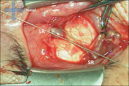
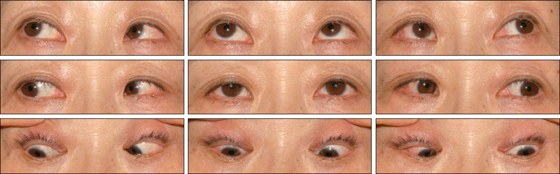
A 51-year-old female patient presented with progressive esotropia and diplopia. According to the Krimsky test, the patient showed 70 prism diopter esotropia and 30 prism diopter hypotropia in her left eye. The axial length was 34.97 mm in the right eye and 33.71 mm in the left eye. The patient was diagnosed with myopic strabismus fixus. The authors performed the Yokoyama procedure on her left eye. Surgical examination revealed each medial rectus muscle was recessed. Half of the muscle bellies of the superior and lateral rectus muscles were sutured together without muscle splitting 15 mm posterior from their insertion. At 1 year postoperatively, the patient showed 30 prism diopter esotropia and 20 prism diopter hypotropia in her right eye by alternative prism cover test. The authors performed the same procedure on her right eye. At 2 months after the second surgery, the patient showed orthotropia in the primary position and gaze limitation was improved.
Go to : 
References
1. Krzizok TH, Schroeder BU. Measurement of recti eye muscle paths by magnetic resonance imaging in highly myopic and normal subjects. Invest Ophthalmol Vis Sci. 1999; 40:2554–60.
2. Mansour AM, Wang F, el-Baba F, Henkind P. Ocular complications in strabismus fixus convergens. Ophthalmologica. 1987; 195:161–6.
3. Basghaw J. The ‘heavy eye' phenomenon. A preliminary report. Br J Ophthalmol. 1966; 23:7.
4. Sturm V, Menke MN, Chaloupka K, Landau K. Surgical treatment of myopic strabismus fixus: a graded approach. Graefes Arch Clin Exp Ophthalmol. 2008; 246:1323–9.

5. Hayashi T, Iwashige H, Maruo T. Clinical features and surgery for acquired progressive esotropia associated with severe myopia. Acta Ophthalmol Scand. 1999; 77:66–71.
6. Demer JL, Von Noorden GK. High myopia as an unusual cause of restrictive motility disturbance. Surv Ophthalmol. 1989; 33:281–4.
7. Yokoyama T, Tabuchi H, Ataka S, et al. The mechanism of development in progressive esotropia with high myopia. Jan-Tjeerd de Faber, editor. Transactions: 26th Meeting, European Strabismological Association, Barcelona, Spain. September 2000. Lisse [Netherlands]: Swets & Zeitlinger;2000. p. 218–21.
8. Krzizok TH, Kaufmann H, Traupe H. Elucidation of restrictive motility in high myopia by magnetic resonance imaging. Arch Ophthalmol. 1997; 115:1019–27.
9. Bagolini B, Tamburrelli C, Dickmann A, Colosimo C. Convergent strabismus fixus in high myopic patients. Doc Ophthalmol. 1990; 74:309–20.
10. Herzau V, Ioannakis K. [Pathogenesis of eso- and hypotropia in high myopia]. Klin Monbl Augenheilkd. 1996; 208:33–6.
11. Demer JL, Miller JM, Poukens V, et al. Evidence for fibromuscular pulleys of the recti extraocular muscles. Invest Ophthalmol Vis Sci. 1995; 36:1125–36.
12. Yamaguchi M, Yokoyama T, Shiraki K. Surgical Procedure for correcting globe dislocation in highly myopic strabismus. Am J Ophthalmol. 2010; 149:341–6.
13. Rutar T, Demer JL. Heavy Eye syndrome in the absence of high myopia: A connective tissue degeneration in elderly strabismic patients. J AAPOS. 2009; 13:36–44.
14. Sharma P, Gupta NK, Arora R, Prakash P. Strabismus fixus convergens secondary to amyloidosis. J Pediatr Ophthalmol Strabismus. 1991; 28:236–7.
15. Nakagawa S, Kii T, Suzuki J, et al. 5 cases of strabismus fixus. Nippon Ganka Kiyo. 1989; 40:656–62.
16. Krzizok TH, Kaufmann H, Traupe H. New approach in strabismus surgery in high myopia. Br J Ophthalmol. 1997; 81:625–30.
17. Larsen PC, Gole GA. Partial Jensen's procedure for the treatment of myopic strabismus fixus. J AAPOS. 2004; 8:393–5.
18. Rowe FJ, Noonan CP. Surgical treatment for progressive esotropia in the setting of high-axial myopia. J AAPOS. 2006; 10:596–7.
19. Wong I, Leo SW, Khoo BK. Loop myopexy for treatment of myopic strabismus fixus. J AAPOS. 2005; 9:589–91.
20. Lee JS, Heo H, Park SW, Park YG. A case of rectus muscle transposition with silicone band in strabismus fixus with high myopia. J Korean Ophthalmol Soc. 2009; 50:1128–32.
21. Durian JM, Maddula S, Marsh IB. Treatment of heavy eye syndrome using simple loop myopexy. J AAPOS. 2010; 14:39–41.
Go to :
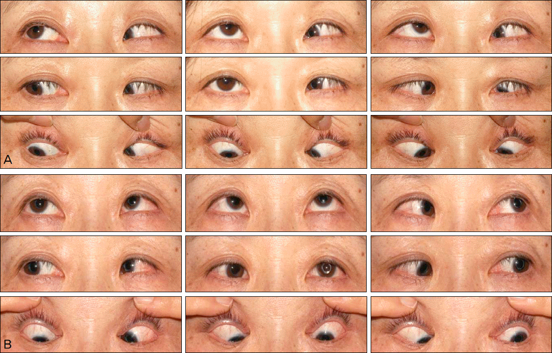 | Figure 1.(A) Preoperative photograph shows limitation of abduction and supraduction of the left eye. In primary position, She showed 70 prism diopter LET and 30 prism diopter LHoT. (B) Postoperative 1 year photograph shows improved movement of left eye. Her preferred eye changed from right to left eye. She showed 30-prism diopter RET and 20 prism diopter RHoT. LET = left esotropia; LHoT = left hypotropia. |
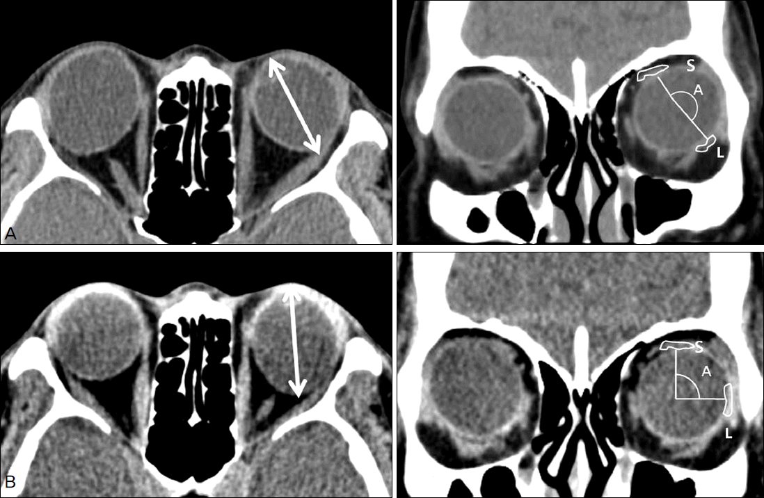 | Figure 2.(A) Preoperative computed tomography shows elongated globe and disproportion between globe and orbit. Downward displacement of lateral rectus muscle is noted. (B) Postoperative computed tomography shows restoration of dislocated globe and improvement of downward displacement of lateral rectus muscle on left eye. A = angle of dislocation; S = superior rectus muscle; L = lateral rectus muscle. |
 XML Download