

 PDF
PDF ePub
ePub Citation
 Print
Print


Abstract
Purpose
To report a case of abducens nerve palsy after a percutaneous nerve blocking procedure for trigeminal neuralgia.
Case summary
A 35-year-old female complaining of stabbing pain in the right maxillary area 4 months in duration was diagnosed with trigeminal neuralgia at a pain clinic. The patient underwent a percutaneous trigeminal nerve blocking procedure using alcohol at the right maxillary nerve. After the procedure, the patient was referred to an ophthalmologic service for horizontal diplopia and abduction defect of her right eye. Her corrected visual acuity, intraocular pressure, pupillary response, anterior segment and fundus were normal bilaterally. The patient had right esotropia of 38 prism diopters in primary gaze (70 prism diopters in right gaze, 20 prism diopters in left gaze) with limited abduction of -3 in the right eye. She was diagnosed with abducens nerve palsy of the right eye. Three months after initial presentation, the patient had intermittent esotropia of 4 prism diopters at right gaze and orthophoria at the other diagnostic gazes; she presented no diplopia.
Conclusions
In the present case study, abducens nerve palsy following a percutaneous trigeminal nerve blocking procedure resolved over 3 months. Because the abducens nerve is adjacent to the trigeminal nerve near the foramen ovale based on anatomical structure, when performing a percutaneous trigeminal blocking procedure, the surgeon should be aware that deep needle puncture could cause abducens nerve palsy.
References
1. Han KR, Kim YS, Kim C. Clinical features of trigeminal neuralgia. Korean J Pain. 2007; 20:174–80.

2. McLeod NM, Patton DW. Peripheral alcohol injections in the management of trigeminal neuralgia. Oral Surg Oral Med Oral Pathol Oral Radiol Endod. 2007; 104:12–7.
3. Peters G, Nurmikko TJ. Peripheral and gasserian ganglion-level procedures for the treatment of trigeminal neuralgia. Clin J Pain. 2002; 18:28–34.
4. Kaplan M, Erol FS, Ozveren MF, et al. Review of complications due to foramen ovale puncture. J Clin Neurosci. 2007; 14:563–8.
5. Kim C, Lee HK, Yang SK, et al. Alcohol block in the treatment of trigeminal neuralgia: a retrospective study to assess its efficacy. J Korean Pain Soc. 1996; 9:83–8.
6. Han KR, Kim C, Kim DW, et al. Long-term outcome of trigeminal nerve block with alcohol for the treatment of trigeminal neuralgia. Korean J Pain. 2006; 19:45–50.
7. Choi YS, Kim YC, Park SH, et al. Percutaneous radiofrequency thermocoagulation for trigeminal neuralgia. Korean J Anesthesiol. 2008; 54:552–6.
8. Jeon C, Sa HS, Oh SY. Causes and natural course of the sixth cranial nerve palsy. J Korean Ophthalmol Soc. 2006; 47:1776–80.
9. Rush JA, Younge BR. Paralysis of cranial nerves III, IV, and VI. Cause and prognosis in 1,000 cases. Arch Ophthalmol. 1981; 99:76–9.
10. Holmes JM, Droste PJ, Beck RW. The natural history of acute traumatic sixth nerve palsy or paresis. J AAPOS. 1998; 2:265–8.
11. Holmes JM, Beck RW, Kip KE, et al. Botulinum toxin treatment versus conservative management in acute traumatic sixth nerve palsy or paresis. J AAPOS. 2000; 4:145–9.
12. Wilkinson HA. Trigeminal nerve peripheral branch phenol/glycerol injections for tic douloureux. J Neurosurg. 1999; 90:828–32.
Figure 1.
(A) Performing percutaneous injection of the nerve V2 through the foramen ovale of the skull base. Coronal C-arm images before (B) and after penetrating ganglion (C). A white dotted oval circle represents the foramen ovale and black arrow heads represent a cannula for lesions.
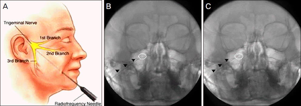
Figure 2.
(A) A 35-year-old female at the onset of right abducens nerve palsy with an abduction deficit of the right eye after blockage procedure. (B) Hess screen test reveals 15 degree angle of subjective esodeviation.
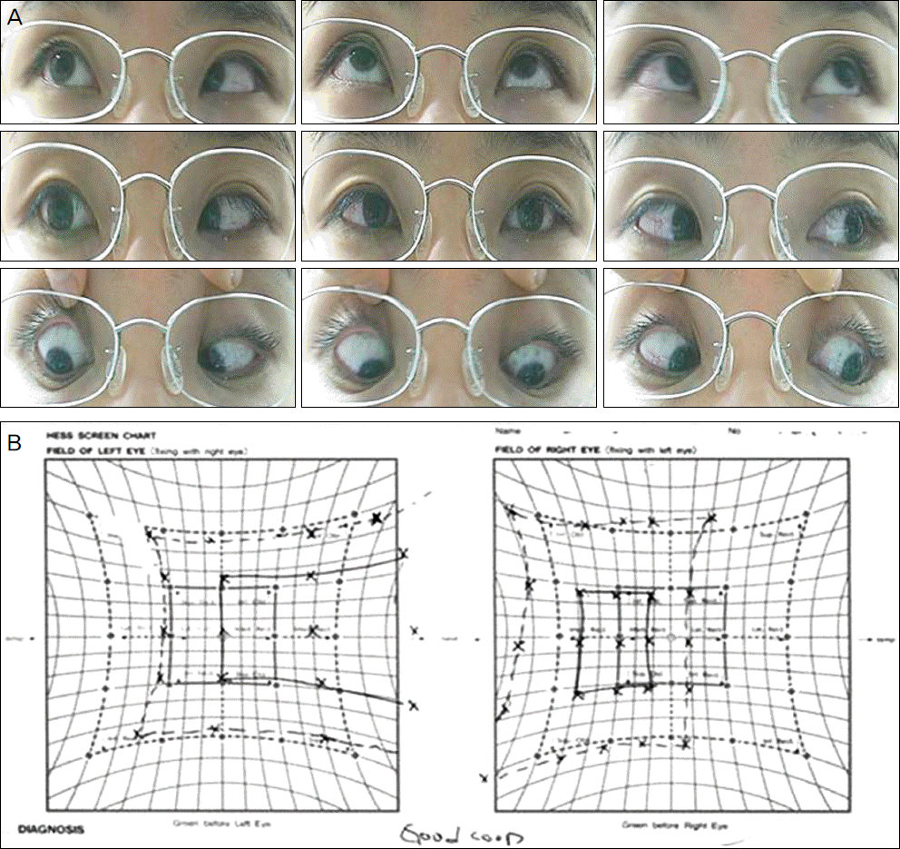
Figure 3.
(A) Diagnostic gaze positions 3 months after initial presentation. Full range of versions in both eyes is noted. (B) Hess screeen test, however, shows small angle of esotropia remains.
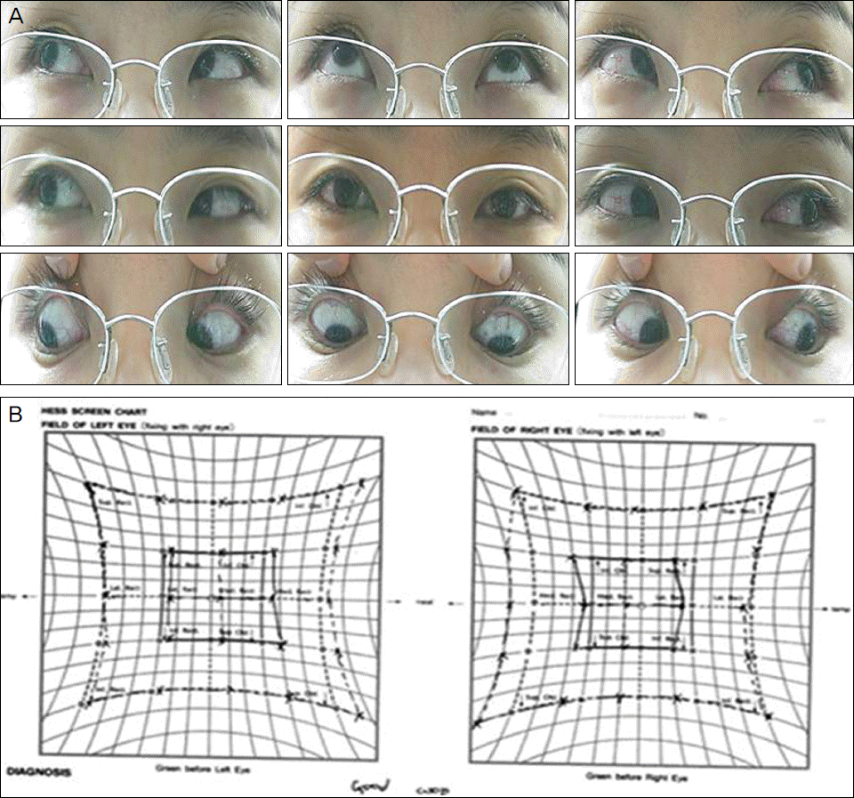
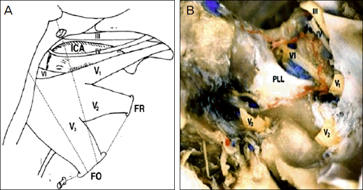
Figure 4.
(A) and (B) shows anatomical relationship between the abducens nerve and trigeminal nerve. FO = foramen ovale; V = trigeminal nerve; VI = abducens nerve; PLL = petrolingual ligament. (Modified from Kaplan M, Erol FS, Ozveren M F, et al. Review of complications due to foramen ovale puncture. J Clin Neurosci 2007;14:563-8)4
 XML Download