

 PDF
PDF ePub
ePub Citation
 Print
Print


Abstract
Purpose
To report a case of severe vaso-occlusive retinopathy with significant decrease of bilateral visual acuity as the first manifestation associated with systemic lupus erythematosus (SLE).
Case summary
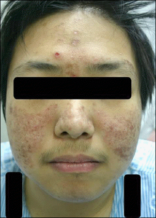
A 23-year-old man was referred to our clinic with bilateral visual impairment of hand motion (HH). Fundus examination revealed severe retinal hemorrhage, cotton-wool patch, occlusive retinal vasculitis with vascular engorgement, and diffuse retinal edema in both eyes. Because of a malar rash on both cheeks, generalized edema was observed on initial examination with hypertension, azotemia, anemia, and thrombocytopenia, The patient was diagnosed with SLE, strongly positive to antinuclear antibody (ANA), and received an intravitreal injection of Bevacizumab (Avastin, Genentech Inc., San Francisco, CA, USA) in the left eye in addition to hemodialysis, transfusion, systemic corticosteroid and immunosuppressant treatment due to lupus nephritis. Eighteen months later, the retinal edema, cotton-wool patch and hemorrhage resolved, leaving epiretinal membrane without traction in his left eye and diffuse degeneration of the right eye. Final visual acuity was HM in the right eye and 20/100 in the left eye.
Go to : 
References
1. Gold DH, Morris DA, Henkind P. Ocular findings in systemic lupus erythematosus. Br J Ophthalmol. 1972; 56:800–4.

2. Dougal MA, Evans LS, McClellan KR, Robinson J. Central retinal artery occlusion in systemic lupus erythematosus. Ann Ophthalmol. 1983; 15:38–40.
3. Hall S, Buettner H, Luthra HS. Occlusive retinal vascular disease in systemic lupus erythematosus. J Rheumatol. 1984; 11:846–50.
4. Graham EM, Spalton DJ, Barnard RO, et al. Cerebral and retinal vascular changes in systemic lupus erythematosus. Ophthalmology. 1985; 92:444–8.
5. Jabs DA, Fine SL, Hochberg MC, et al. Severe retinal vaso-occlusive disease in systemic lupus erythematous. Arch Ophthalmol. 1986; 104:558–63.
6. Au A, O'Day J. Review of severe vaso-occlusive retinopathy in systemic lupus erythematosus and the antiphospholipid syndrome: associations, visual outcomes, complications and treatment. Clin Experiment Ophthalmol. 2004; 32:87–100.
7. Giorgi D, Pace F, Giorgi A, et al. Retinopathy in systemic lupus erythematosus: pathogenesis and approach to therapy. Hum Immunol. 1999; 60:688–96.
8. Klinkhoff AV, Beattie CW, Chalmers A. Retinopathy in systemic lupus erythematosus: relationship to disease activity. Arthritis Rheum. 1986; 29:1152–6.
9. Yoon CK, Park JH, Yu HG. Retinopathy associated with systemic lupus erythematosus. J Korean J Ophthalmol Soc. 2009; 50:1215–20.
10. Quismorio FP Jr.Clinical application of serologic abnormalities in systemic lupus erythematosus. In : Wallace BJ, Hahn BH, editors. Dubois' Lupus Erythematosus. 5th ed.Baltimore: Lippincott Williams & Wilkins;1997. p. 925–42.
11. Mills JA. Systemic lupus erythematosus. N Engl J Med. 1994; 330:1871–9.
12. Stafford-Brady FJ, Urowitz MB, Gladman DD, Easterbrook M. Lupus retinopathy. Patterns, associations, and prognosis. Arthritis Rheum. 1988; 31:1105–10.
13. Ingram SB, Goodnight SH Jr., Bennett RM. An unusual syndrome of a devastating non-inflammatory vasculopathy associated with anticardiolipin antibodies: report of two cases. Arthritis Rheum. 1987; 30:1167–72.
14. Montehermoso A, Cervera R, Font J, et al. Association of anti-phospholipid antibodies with retinal vascular disease in systemic lupus erythematosus. Semin Arthritis Rheum. 1999; 28:326–32.
15. Asherson RA, Merry P, Acheson JF, et al. Antiphospholipid antibodies: a risk factor for occlusive ocular vascular disease in systemic lupus erythematosus and the ‘primary' antiphospholipid syndrome. Ann Rheum Dis. 1989; 48:358–61.
16. Fitzpatrick EP, Chesen N, Rahn EK. The lupus anticoagulant and retinal vaso-occlusive disease. Ann Ophthalmol. 1990; 22:148–52.
17. Nag TC, Wadhwa S. Vascular changes of the retina and choroid in systemic lupus erythematosus: pathology and pathogenesis. Curr Neurovasc Res. 2006; 3:159–68.
18. Gold D, Feiner L, Henkind P. Retinal arterial occlusive disease in systemic lupus erythematosus. Arch Ophthalmol. 1977; 95:1580–5.
19. Ushiyama O, Ushiyama K, Koarada S, et al. Retinal disease in patients with systemic lupus erythematosus. Ann Rheum Dis. 2000; 59:705–8.
20. Kleiner RC, Najarian LV, Schatten S, et al. Vaso-occlusive retinopathy associated with antiphospholipid antibodies (lupus anticoagulant retinopathy). Ophthalmology. 1989; 96:896–904.
21. Peponis V, Kyttaris VC, Tyradellis C, et al. Ocular manifestations of systemic lupus erythematosus: a clinical review. Lupus. 2006; 15:3–12.
22. Kuryliszyn-Moskal A, Klimiuk PA, Sierakowski S, Ciolkiewicz M. Vascular endothelial growth factor in systemic lupus erythematosus: relationship to disease activity, systemic organ manifestation, and nailfold capillaroscopic abnormalities. Arch Immunol Ther Exp. 2007; 55:179–85.
23. Kurup S, Lew J, Byrnes G, et al. Therapeutic efficacy of intravitreal bevacizumab on posterior uveitis complicated by neovascularization. Acta Ophthalmol. 2009; 87:349–52.
24. Lee WJ, Cho HY, Lee YJ, et al. Intravitreal bevacizumab for severe vaso-occlusive retinopathy in systemic lupus erythematosus. Rheumatol Int. 2011; Sep 28.
25. Jung NH, Kim SY. A case of severe retinal vaso-occlusive disease in systemic lupus erythematosus. J Korean Ophthalmol Soc. 1993; 34:1287–92.
Go to :
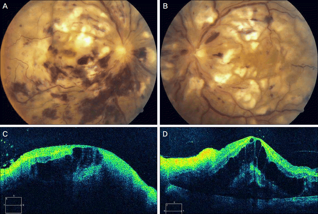 | Figure 1.Fundus (upper) and optical coherence tomographic (lower) findings of the right (A and C) and left (B and D) eyes at presentation. (A, B) Fundus findings revealed vessel engorgement, multiple flame-shaped retinal hemorrhages, cotton wool patchs, diffuse retinal edema and optic disc swelling. (C, D) Spectal-domein optical coherence tomographic findings showed severe macular cystoids edema (both eyes) and serous retinal detachment (left eye). |
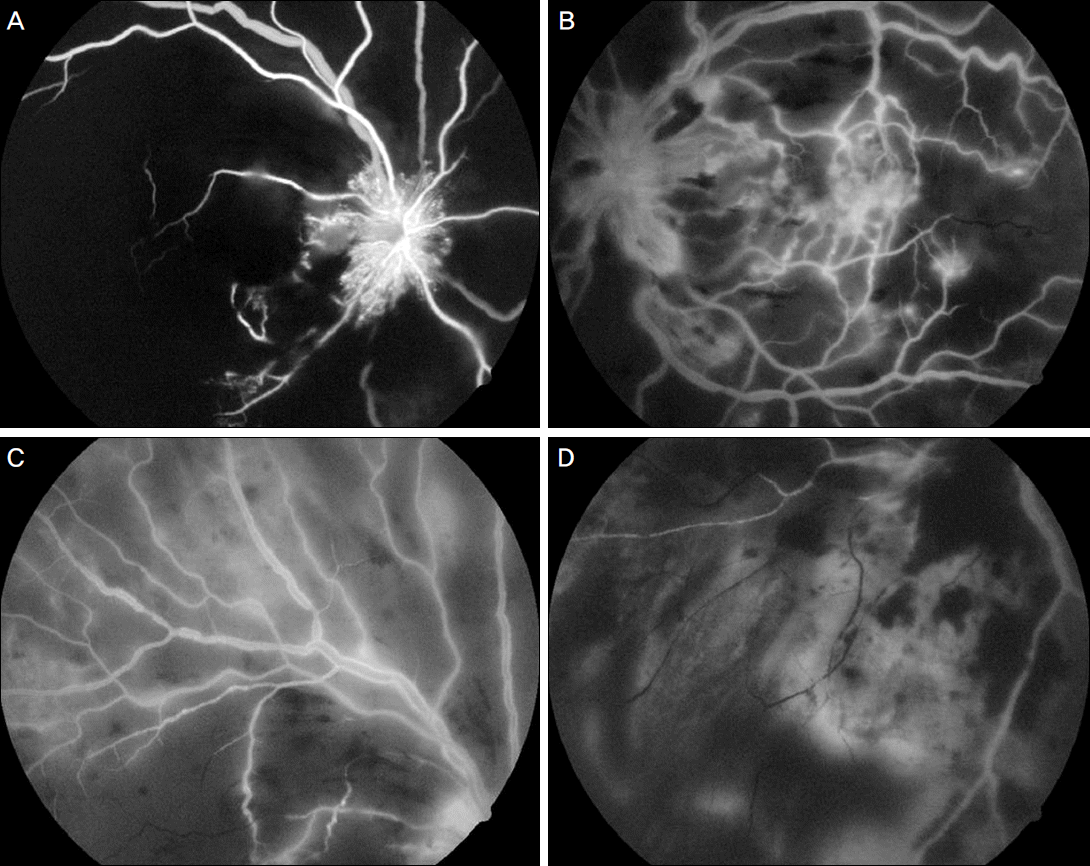 | Figure 3.Fluorescein angiographic findings showed neovascularization at the optic dsc, total defect of choroidal filling, occlusion of the inferior retinal artery, diffuse leakage around the vessel of the right eye (A and C), and leakage around the optic disc head, diffuse peripheral non-perfusion area, multiple occlusions of peripheral retinal vessels (B and D). |
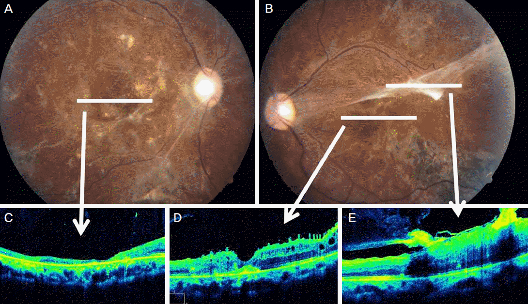 | Figure 4.Final ocular findings of the right (A and C) and left (B, D and E) eyes. (A, C) Fundus photographs show diffuse degenerative changes, pale optic disc, narrowing of arteries, and OCT findings revealed diffuse retinal thinning and uncertain foveal contour. (B, D, E) fundus photograph show focal degenerative change and parafoveal tractional fibrotic membrane, and OCT findings reveals relatively clear foveal contour and parafoveal, focal retinal detachment due to tractional membrane. |
 XML Download