

 PDF
PDF ePub
ePub Citation
 Print
Print


Abstract
Purpose
To introduce a case of complicated ophthalmopathy in herpes zoster ophthalmicus including vitreous opacity, retinal hemorrhage and optic neuropathy having components of anterior ischemic optic neuropathy and optic neuritis.
Case summary
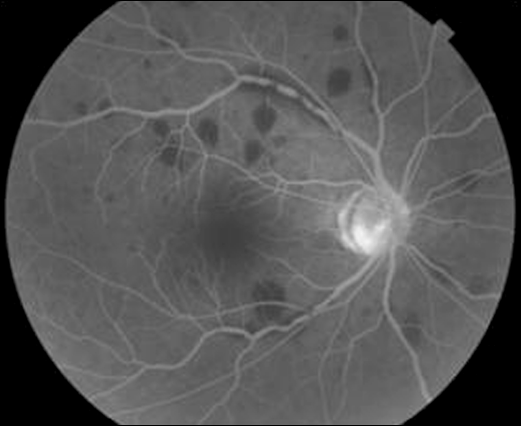
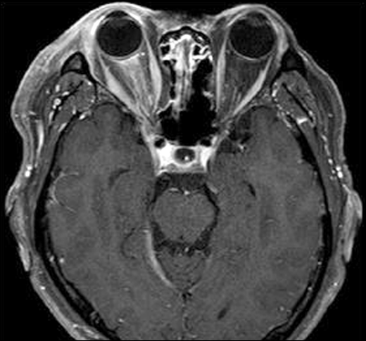
A 59-year-old man visited our clinic because of visual disturbance in the right eye which occurred after right facial pain and vesicles. There were inflammatory cells in the anterior chamber, retinal hemorrhage in the retina and vitreous opacity was found. Track-like high signal intensity along the right optic nerve was found on T1 MRI. Partial filling defect of optic disc was observed on fluorescein angiography (FAG). The patient was diagnosed with herpes zoster ophthalmicus complicated by anterior uveitis and optic neuropathy having components of anterior ischemic optic neuropathy and optic neuritis. The patient was started on intravenous acyclovir at a dose of 10 mg/kg every 8 hours for 5 days and Herpesid eye ointment 5 times daily. After the initial treatment, oral acyclovir 400 mg was given 3 times daily for 14 days. Skin symptoms and fundus findings improved but the visual acuity did not improve because of optic atrophy.
Go to : 
References
1. The Korean External Eye Disease Society. Cornea. 2nd ed.Ilchokak;2005. p. 127–70.
2. Lee HJ, Kim SY, Jung MS. The clinical characteristics of facial herpes zoster in Korean patients. J Korean Ophthalmol Soc. 2010; 51:8–13.

3. Lee HR, Cho BC. A clinical study of herpes zoster ophthalmicus. J Korean Ophthalmol Soc. 1988; 29:387–91.
4. Kim JY, Ahn M, Lee DW. Two cases of optic neuritis in herpes zoster ophthalmicus. J Korean Ophthalmol Soc. 2008; 49:1028–32.
5. Zaal MJ, Völker-Dieben HJ, D'Amaro J. Visual prognosis in immunocompetent patients with herpes zoster ophthalmicus. Acta Ophthalmol Scand. 2003; 81:216–20.
6. Park SH, Kim WJ, Yang SW, Kim MS. Herpes zoster ophthalmicus complicated by hyphema, glaucoma and external ophthalmoplegia. J Korean Ophthalmol Soc. 2007; 48:1573–8.
7. Wang TJ, Hu CC, Lin HC. Increased risk of anterior uveitis following herpes zoster: a nationwide population-based study. Arch Ophthalmol. 2012; 130:451–5.
8. Bjerrum SS, Hessellund A. [Complete ophthalmoplegia following outburst of herpes zoster]. Ugeskr Laeger. 2012; 174:1832–3.
9. Hesse RJ. Herpes zoster ophthalmicus associated with delayed retinal thrombophlebitis. Am J Ophthalmol. 1977; 84:329–31.
10. Borruat FX, Herbort CP. Herpes zoster ophthalmicus. Anterior ischemic optic neuropathy and acyclovir. J Clin Neuroophthalmol. 1992; 12:37–40.
11. Liesegang TJ. Herpes zoster ophthalmicus natural history, risk factors, clinical presentation, and morbidity. Ophthalmology. 2008; 115((2 Suppl)):S3–12.
12. Lexa FJ, Galetta SL, Yousem DM, et al. Herpes zoster ophthalmicus with orbital pseudotumor syndrome complicated by optic nerve infarction and cerebral granulomatous angiitis: MRpathologic correlation. AJNR Am J Neuroradiol. 1993; 14:185–90.
13. Beck RW, Trobe JD. The Optic Neuritis Treatment Trial. Putting the results in perspective. The Optic Neuritis Study Group. J Neuroophthalmol. 1995; 15:131–5.
14. Wood MJ, Johnson RW, McKendrick MW, et al. A randomized trial of acyclovir for 7 days or 21 days with and without prednisolone for treatment of acute herpes zoster. N Engl J Med. 1994; 330:896–900.
Go to :
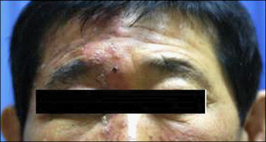 | Figure 1.Skin eruptions corresponding to the dermatome of the right ophthalmic branch of the trigerminal nerve are seen. |
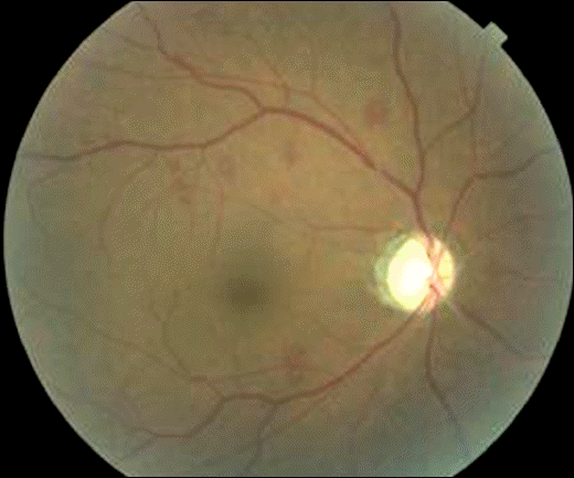
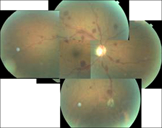 | Figure 2.Multiple round-shaped retinal hemorrhage, flame-shaped retinal hemorrhage and vitreous opacity are seen. |
 XML Download