

 PDF
PDF ePub
ePub Citation
 Print
Print


Abstract
Purpose
To investigate clinical characteristics of post-traumatic intraocular foreign body (IOFB), which occurred between 2006 and 2010, and prognostic factors associated with final visual outcome.
Methods
A retrospective chart review was performed of patients with IOFB who visited our clinic from January 1, 2006 to December 31, 2010, and who were followed up for more than 6 months. Cross tabulation and correlation analyses were conducted to evaluate the predictive factors related to final visual acuity.
Results
A total of 36 eyes (35 patients) were included in this study. The mean age was 44.2 years, and 33 patients (94.3%) were male. The most common cause of IOFB was lawnmower-related activity (12 patients; 33.3%) and hammering-related activity (11 patients; 30.1%). Among analyzed prognostic factors, only initial visual acuity was significantly correlated with visual outcomes (p < 0.001).
Conclusions
Compared with previous studies, lawnmower and hammering-related activities were still the most common causes of IOFB, however, the incidence of occurrence in the 40's and 50's was relatively higher than in previous studies. Considering the poor visual outcome of IOFB even after proper surgical treatment, using proper eye protection should be emphasized in a dangerous work environment to prevent ocular trauma.
Go to : 
References
1. Shock JP, Adams D. Long-term visual acuity results after penetrating and perforating ocular injuries. Am J Ophthalmol. 1985; 100:714–8.

2. Kim JY, Kim JW, Lee J. Clinical evaluations of penetration ocular injuries. J Korean Ophthalmol Soc. 1992; 33:919–24.
3. Mines M, Thach A, Mallonee S, et al. Ocular injuries sustained by survivors of the Oklahoma City bombing. Ophthalmology. 2000; 107:837–43.
4. Kim JH, Yang SJ, Kim DS, et al. Fourteen-year review of open globe injuries in an urban Korean population. J Trauma. 2007; 62:746–9.
5. Lim HS, Hahn DK. A reiew of intraocular foreignbodies. J Korean Ophthalmol Soc. 1991; 32:975–83.
6. Park CH, Oum BS. A clinical evaluation of intraocular foreign bodies. J Korean Ophthalmol Soc. 1991; 32:498–508.
7. Cho HJ, Seo MS. Intraocular foreign bodies: clinical characteristics and visual prognosis. J Korean Ophthalmol Soc. 2002; 43:1968–75.
8. Williams DF, Mieler WF, Abrams GW, Lewis H. Results and prognostic factors in penetrating ocular injuries with retained intraocular foreign bodies. Ophthalmology. 1988; 95:911–6.
9. De Souza S, Howcroft MJ. Management of posterior segment intraocular foreign bodies: 14 years' experience. Can J Ophthalmol. 1999; 34:23–9.
10. Camacho H, Mejia LF. Extraction of intraocular foreign bodies by pars plana vitrectomy. A retrospective study. Ophthalmologica. 1991; 202:173–9.
11. Elder MJ. Penetrating eye injuries in children of the West Bank and Gaza strip. Eye (Lond). 1993; 7:429–32.
12. Zhang Y, Zhang M, Jiang C, Qiu HY. Intraocular foreign bodies in china: clinical characteristics, prognostic factors, and visual outcomes in 1,421 eyes. Am J Ophthalmol. 2011; 152:66–73.
13. Kim JM, Kim CW. Factors influencing the prognosis of corneoscleral laceration. J Korean Ophthalmol Soc. 1985; 26:311–9.
14. Lee EH, Moon CS, Lee SY, Lew HM. Factors influencing final visual outcome in intraocular foreign bodies. J Korean Ophthalmol Soc. 2001; 42:997–1002.
15. Jang SG, Lee SJ. Statistical evaluation for perforating ocular injuries. J Korean Ophthalmol Soc. 1988; 29:921–9.
16. Kim HJ, Kwon JY. A clinical observation of perforating ocular injuries. J Korean Ophthalmol Soc. 1989; 30:123–30.
17. Kim SY, Hahn DK. Clinical evaluation of the retinal injuries following perforating ocular traumas. J Korean Ophthalmol Soc. 1995; 36:1171–8.
18. Hutton WL, Fuller DG. Factors influencing final visual results in severely injured eyes. Am J Ophthalmol. 1984; 97:715–22.
19. Jeong JY, Park YK, Kim SD, Kim TW. The postoperative results and the risk factors between removed and retained intraocular foreign bodies. J Korean Ophthalmol Soc. 2004; 45:425–37.
20. Greven CM, Engelbrecht NE, Slusher MM, Nagy SS. Intraocular foreign bodies: management, prognostic factors, and visual outcomes. Ophthalmology. 2000; 107:608–12.
21. Chiquet C, Zech JC, Denis P, et al. Intraocular foreign bodies. Factors influencing final visual outcome. Acta Ophthalmol Scand. 1999; 77:321–5.
22. Chiquet C, Zech JC, Gain P, et al. Visual outcome and prognostic factors after magnetic extraction of posterior segment foreign bodies in 40 cases. Br J Ophthalmol. 1998; 82:801–6.
23. Punnonen E, Laatikainen L. Prognosis of perforating eye injuries with intraocular foreign bodies. Acta Ophthalmol (Copenh). 1989; 67:483–91.
24. Coleman DJ, Lucas BC, Rondeau MJ, Chang S. Management of intraocular foreign bodies. Ophthalmology. 1987; 94:1647–53.
25. Jonas JB, Budde WM. Early versus late removal of retained intraocular foreign bodies. Retina. 1999; 19:193–7.
26. Wani VB, Al-Ajmi M, Thalib L, et al. Vitrectomy for posterior segment intraocular foreign bodies: visual results and prognostic factors. Retina. 2003; 23:654–60.
Go to :
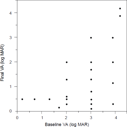 | Figure 1.Correlation between initial visual acuity and final visual acuity. Visual acuity was converted to log MAR (R = 0.644, p-value: < 0.001). VA = visual acuity. |
Table 1.
Demographic features
Table 2.
Characteristics and oular findings in eyes with intraocular foreign body (n = 36)
Table 3.
Prognostic factors associated with poor visual outcome (visual acuity, <Snellen, 0.1)
| Factors | Number (% in total) | Final VA (<Snellen, 0.1) Total No. (% in row) | p-value | |
|---|---|---|---|---|
| Property | ||||
| Metal (Magnetic) | 22 (61.1) | 13 (59.1) | 0.86* | |
| Metal (Non-magnetic) | 2 (5.6) | 1 (50.0) | ||
| Non-metal | 12 (33.3) | 6 (50.0) | ||
| Location | ||||
| Anterior | 10 (27.8) | 3 (30.0) | 0.07* | |
| Posterior | 26 (72.2) | 17 (65.4) | ||
| Inlet | ||||
| Cornea | 28 (77.8) | 17 (60.7) | 0.42* | |
| Sclera | 8 (22.2) | 3 (37.5) | ||
| Limbus | 0 (0) | 0 (0) | ||
| Length of IOFB | ||||
| <3 mm | 23 (63.9) | 10 (43.5) | 0.08* | |
| ≥3 mm | 13 (36.1) | 10 (76.9) | ||
| Timing of IOFB removal | ||||
| ≤48 hr | 31 (86.1) | 17 (54.8) | 1.00* | |
| >48 hr | 5 (13.9) | 3 (60.0) | ||
| Ocular findings | ||||
| Hyphema | 25 (69.4) | 16 (64.0) | 0.16* | |
| Iris damage | 21 (58.3) | 14 (66.7) | 0.11† | |
| Traumatic cataract | 26 (72.2) | 17 (65.3) | 0.07* | |
| Vitreous hemorrhage | 22 (61.1) | 13 (59.1) | 0.59† | |
| Endophthalmitis | 8 (22.2) | 5 (62.5) | 0.71* | |
| Retinal tear | 8 (22.2) | 7 (87.5) | 0.05* |
 XML Download