

 PDF
PDF ePub
ePub Citation
 Print
Print


Abstract
Purpose
To compare the efficacy and intraoperative characteristics of DisCoVisc with those of Hyal 2000 (sodium hyaluronate 1.0%) in cataract surgery.
Methods
Cataract surgery was performed on 60 eyes in 49 patients who were diagnosed with moderate cataracts. 30 eyes were performed with DisCoVisc and a control group with 30 eyes using Hyal 2000 (sodium hyaluronate 1.0%). Phacodynamics was evaluated including ultrasound (US) time, mean US intensity, cumulative dissipated energy (CDE), and amount of used balanced salt solution. Corneal endothelium and corneal thickness were measured preoperatively and 1 day and 1 month and 2 months postoperatively.
Results
There were no statistically significant differences in phacodynamic parameters in the two groups. The central cor-neal thickness change from preoperatively to postoperatively in the DisCoVisc group was +0.07 ± 2.44 μm and Hyal 2000 group +0.84 ± 2.93 μm (p = 0.032) at 2 months. Corneal endothelial cell loss (ECL)(%) at 2 months was 7.67 ± 8.01% in DisCoVisc group and 13.23 ± 15.5% in the Hyal 2000 group (p = 0.005).
Go to : 
References
1. Storr-Paulsen A, Norregaard JC, Ahmed S, et al. Endothelial cell damage after cataract surgery: divide-and-conquer versus phaco-chop technique. J Cataract Refract Surg. 2008; 34:996–1000.

2. Fishkind W, Bakewell B, Donnenfeld ED, et al. Comparative clinical trial of ultrasound phacoemulsification with and without the WhiteStar system. J Cataract Refract Surg. 2006; 32:45–9.
3. Hoffman RS, Fine IH, Packer M. New phacoemulsification technology. Curr Opin Ophthalmol. 2005; 16:38–43.
4. Madsen K, Steveni U, Apple DJ, et al. Histochemical and receptor binding studies of hyaluronic acid binding sites on the corneal endothelium. Ophthalmic Pract. 1989; 7:92–7.
5. Arshinoff SA. Dispersive-cohesive viscoelastic soft shell technique. J Cataract Refract Surg. 1999; 25:167–73.
6. Rainer G, Schmid KE, Findl O, et al. Natural course of intraocular pressure after cataract surgery with sodium hyaluronate 1% versus hydroxypropylmethylcellulose 2%. Ophthalmology. 2007; 114:1089–93.
7. Petroll WM, Jafari M, Lane SS, et al. Quantitative assessment of ophthalmic viscosurgical device retention using in vivo confocal microscopy. J Cataract Refract Surg. 2005; 31:2363–8.
8. Dick HB, Schwenn O. Viscoelastics in ophthalmic surgery. New York: Springer Verlag;2000. p. 7–24.
9. Holzer MP, Tetz MR, Auffarth GU, et al. Effect of Healon5 and 4 other viscoelastic substances on intraocular pressure and endothelium after cataract surgery. J Cataract Refract Surg. 2001; 27:213–8.
10. Kim H, Joo CK. Efficacy of the soft-shell technique using Viscoat and Hyal-2000. J Cataract Refract Surg. 2004; 30:2366–70.
11. Binder PS, Sternberg H, Wickman MG, Worthen DM. Corneal endothelial damage associated with phacoemulsification. Am J Ophthalmol. 1976; 82:48–54.
12. Petroll WM, Jafari M, Lane SS, et al. Quantitative assessment of ophthalmic viscosurgical device retention using in vivo confocal microscopy. J Cataract Refract Surg. 2005; 31:2363–8.
Go to :
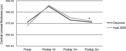 | Figure 1.Change of preoperative and postpoerative central corneal thickness (μm).
p = 0.032. |
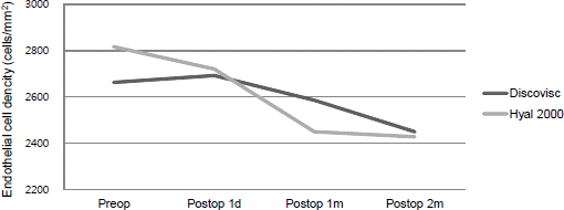 | Figure 2 .Change of preoperative and postoperative corneal endothelial cell density (cells/mm2). |
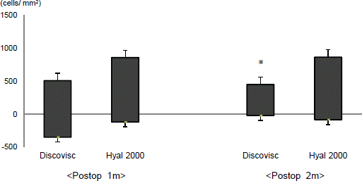 | Figure 3.Endothelial cell loss between preoperation and follow up 1 m, 2 m. *Endothelial cell loss rate follow up 2 m (p = 0.005). |
Table 1.
Preoperative parameters
Table 2.
Intraoperative characteristics of the parameters
 XML Download