Abstract
Purpose
To evaluate the changes in tearfilm, corneal sensation and ocular surface after advanced surface ablation.
Methods
Tearfilm break-up time (BUT), Schirmer test without local anesthesia, fluorescein staining, corneal sensitivity test, ocular surface disease index (OSDI), and conjunctival impression cytology were evaluated in 50 eyes of 25 patients who underwent advanced surface ablation preoperatively and postoperatively at 2 weeks and at 1, 2, 3, and 6 months. Each value was compared to the preoperative value.
Results
OSDI diminished by 2 weeks postoperatively, and corneal sensation diminished by 1 month postoperatively (p < 0.05). There were significant decreases in BUT by 2 weeks to 1 month postoperatively as well as decreases in the Schirmer test by 2 to 3 months postoperatively (p < 0.05). Fluorescein staining increased at 2 weeks postoperatively (p <0.05). Goblet cells decreased substantially by 1 month postoperatively and conjunctival squamous metaplasia increased significantly by 2 months postoperatively (p < 0.05).
Go to : 
References
1. Shin KH, Shyn KH. 2007 survey for KSCRS members: current trends in refractive surgery in Korea. J Korean Ophthalmol Soc. 2009; 50:1468–74.
2. Trattler WB, Barnes SD. Current trends in advanced surface ablation. Curr Opin Ophthalmol. 2008; 19:330–4.

3. Ghadhfan F, Al-Rajhi A, Wagoner MD. Laser in situ keratomileusis versus surface ablation: visual outcomes and complications. J Cataract Refract Surg. 2007; 33:2041–8.

4. Lee SB, Chung MS. Advanced Surface Ablation-Photorefractive Keratectomy (ASA-PRK): Safety and clinical outcome for the correction of mild to moderate myopia with a thin cornea. J Korean Ophthalmol Soc. 2006; 47:1274–86.
5. Ang RT, Dartt DA, Tsubota K. Dry eye after refractive surgery. Curr Opin Ophthalmol. 2001; 12:318–22.

6. Hong JW, Kim HM. The changes of tear break up time after myopic excimer laser photorefractive keratectomy. Korean J Ophthalmol. 1997; 11:89–93.

7. Lee JB, Ryu CH, Kim J, et al. Comparison of tear secretion and tear film instability after photorefractive keratectomy and laser in situ keratomileusis. J Cataract Refract Surg. 2000; 26:1326–31.

8. Albietz JM, McLennan SG, Lenton LM. Ocular surface management of photorefractive keratectomy and laser in situ keratomileusis. J Refract Surg. 2003; 19:636–44.

9. Bron AJ, Evans VE, Smith JA. Grading of corneal and conjunctival staining in the context of other dry eye tests. Cornea. 2003; 22:640–50.

11. Albietz JM, Bruce AS. The conjunctival epithelium in dry eye sub-types: effect of preserved and non-preserved topical treatments. Curr Eye Res. 2001; 22:8–18.

12. Stern ME, Beuerman RW, Fox RI, et al. The pathology of dry eye: the interaction between the ocular surface and lacrimal glands. Cornea. 1998; 17:584–9.
13. Stern ME, Gao J, Siemasko KF, et al. The role of the lacrimal functional unit in the pathophysiology of dry eye. Exp Eye Res. 2004; 78:409–16.

14. Nakamori K, Odawara M, Nakajima T, et al. Blinking is controlled primarily by ocular surface conditions. Am J Ophthalmol. 1997; 124:24–30.

15. Pérez-Santonja JJ, Sakla HF, Cardona C, et al. Corneal sensitivity after photorefractive keratectomy and laser in situ keratomileusis for low myopia. Am J Ophthalmol. 1999; 127:497–504.

16. Murphy PJ, Corbett MC, O'Brart DP, et al. Loss and recovery of corneal sensitivity following photorefractive keratectomy for myopia. J Refract Surg. 1999; 15:38–45.
17. Campos M, Hertzog L, Garbus JJ, McDonnell PJ. Corneal sensitivity after photorefractive keratectomy. Am J Ophthalmol. 1992; 114:51–4.

18. Lawrenson JG, Corbett MC, O'Brart DP, Marshall J. Effect of beam variables on corneal sensitivity after excimer laser photorefractive keratectomy. Br J Ophthalmol. 1997; 81:686–90.

19. Nejima R, Miyata K, Tanabe T, et al. Corneal barrier function, tear film stability, and corneal sensation after photorefractive keratectomy and laser in situ keratomileusis. Am J Ophthalmol. 2005; 139:64–71.

20. Kauffmann T, Bodanowitz S, Hesse L, Kroll P. Corneal reinnervation after photorefractive keratectomy and laser in situ keratomileusis: an in vivo study with a confocal videomicroscope. Ger J Ophthalmol. 1996; 5:508–12.
21. Erie JC, McLaren JW, Hodge DO, Bourne WM. Recovery of cor-neal subbasal nerve density after PRK and LASIK. Am J Ophthalmol. 2005; 140:1059–64.

22. Horwath-Winter J, Vidic B, Schwantzer G, Schmut O. Early changes in corneal sensation, ocular surface integrity, and tear-film function after laser-assisted subepithelial keratectomy. J Cataract Refract Surg. 2004; 30:2316–21.

23. Herrmann WA, Shah CP, von Mohrenfels CW, et al. Tear film function and corneal sensation in the early postoperative period after LASEK for the correction of myopia. Graefes Arch Clin Exp Ophthalmol. 2005; 243:911–6.

24. Kim MY, Chung SK. The changes of tear break-up time and schirmer's test after photorefrective keratectomy. J Korean Ophthalmol Soc. 2001; 42:228–34.
25. Ozdamar A, Aras C, Karakas N, et al. Changes in the tear flow and tear film stability after photorefractive keratectomy. Cornea. 1999; 18:437–9.
26. Siganos DS, Popescu CN, Siganos CS, Pistola G. Tear secretion following spherical and astigmatic excimer laser photorefractive keratectomy. J Cataract Refract Surg. 2000; 26:1585–9.

27. Battat L, Macri A, Dursun D, Pflugfelder SC. Effects of laser in situ keratomileusis on tear production, clearance, and the ocular surface. Ophthalmology. 2001; 108:1230–5.

28. Wilson SE. Laser in situ keratomileusis-induced (presumed) neurotropic epitheliopathy. Ophthalmology. 2001; 108:1082–7.
29. Egbert PR, Lauber S, Maurice DM. A simple conjunctival biopsy. Am J Ophthalmol. 1977; 84:798–801.

30. Smith RE. The tear film complex: pathogenesis and emerging 414 www.ophthalmology.org therapies for dry eyes. Cornea. 2005; 24:1–7.
31. Johnson ME, Murphy PJ. Changes in the tear film and ocular surface from dry eye syndrome. Prog Retin Eye Res. 2004; 23:449–74.

32. Adams GG, Dilly PN, Kirkness CM. Monitoring ocular disease by impression cytology. Eye (Lond). 1988; 2:506–16.

33. Pflugfelder SC, Tseng SC, Yoshino K, et al. Correlation of goblet cell density and mucosal epithelial membrane mucin expression with rose bengal staining in patients with ocular irritation. Ophthalmology. 1997; 104:223–35.

34. Marner K. ‘Snake-like' appearance of nuclear chromatin in conjunctival epithelial cells from patients with keratoconjunctivitis sicca. Acta Ophthalmol (Copenh). 1980; 58:849–53.

35. Knop E, Brewitt H. Induction of conjunctival epithelial alterations by contact lens wearing. A prospective study. Ger J Ophthalmol. 1992; 1:125–34.
36. Cakmak SS, Unlü MK, Karaca C, et al. Effects of soft contact lenses on conjunctival surface. Eye Contact Lens. 2003; 29:230–3.
Go to : 
 | Figure 1.Impression cytological finding of goblet cell density before and after advanced surface ablation. Photograph exhibits a sharp reduction in goblet cells at 2 weeks post-op, then shows a gradual recovery returning to a pre-op level at between 2-3 months post-op. The increase in goblet cell number continues to 6 months post-op (PAS-H&E, ×100). |
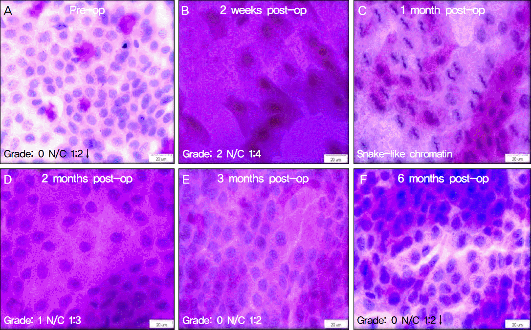
 | Figure 2.Impression cytological finding of squamous metaplasia before and after advanced surface ablation. The photograph shows that epithelial metaplasia grade worsens drastically at two weeks post-op, maximizing at 4 weeks post-op. Then it improves gradu-ally, returning to the pre-op level at 3 months post-op, yet to improve further all the way up to 6 months post-op (PAS-H&E, ×400). |
Table 1.
Demographic features of patients
Table 2.
Clinical parameters before and after
| Parameter | Pre-op | Postoperative | ||||
|---|---|---|---|---|---|---|
| 2 weeks | 1 month | 2 months | 3 months | 6 months | ||
| BUT (seconds) | 6.56 ± 3.14 | 4.60 ± 2.89* | 4.92 ± 2.60* | 5.88 ± 2.60 | 6.24 ± 2.93 | 6.76 ± 2.09 |
| Schirmer I (mm) | 13.6 ± 12.25 | 11.28 ± 9.24 | 10.88 ± 8.14 | 9.16 ± 7.59* | 8.32 ± 9.32* | 9.60 ± 5.35 |
| Corneal fluorescein staining | 0.72 ± 1.21 | 1.6 ± 1.22* | 0.48 ± 0.65 | 0.48 ± 0.71 | 0.36 ± 0.76 | 0.44 ± 0.77 |
| Corneal sensation (mm) | 56.00 ± 4.30 | 50.00 ± 5.20* | 52.00 ± 5.20* | 56.80 ± 2.84 | 57.20 ± 3.84 | 59.00 ± 2.04 |
| OSDI | 29.16 ± 22.71 | 36.86 ± 13.79* | 27.53 ± 13.77 | 25.01 ± 13.13 | 21.25 ± 10.65 | 20.84 ± 12.42 |
Table 3.
The changes in conjunctival impression cytology before and after advanced surface ablation
| Parameter | Pre-op | Postoperative | ||||
|---|---|---|---|---|---|---|
| 2 weeks | 1 month | 2 months | 3 months | 6 months | ||
| Conjunctival epithelial metaplasia | 0.72 ± 0.74 | 1.74 ± 0.56* | 1.82 ± 0.54* | 1.23 ± 0.65* | 0.96 ± 0.69 | 0.51 ± 0.49 |
| Goblet cell count (cells/mm2) | 301.6 ± 205.6 | 102.7 ± 75.1* | 181.0 ± 178.0* | 274.1 ± 148.1 | 353.3 ± 224.1 | 464.5 ± 251.0 |
| Snake‐like chromatin (%) | 32 | 32 | 44 | 40 | 52 | 32 |


 PDF
PDF ePub
ePub Citation
 Print
Print


 XML Download