Abstract
Purpose
The present study assessed the influence of overnight orthokeratology (ortho-k) on the myopic progression in Korean children and analyzed factors affecting myopic progression.
Methods
The ortho-k group was comprised of 31 patients satisfying the inclusion criteria for ortho-k. In the ortho-k group, spherical equivalent refractive error (SER) was measured at baseline, and after 2 weeks, 6 and 12 months. The control group was comprised of 31 patients who were matched according to age, gender, and baseline SER of the ortho-k subjects.
Results
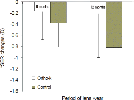
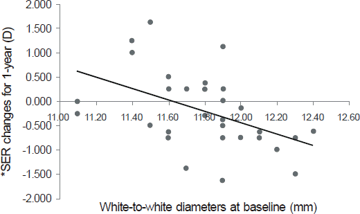
In the ortho-k group, the mean ± SD changes in SER from 2 weeks to 6 months, 6 to 12 months, and 2 weeks to 12 months were -0.17 ± 0.50 D, -0.04 ± 0.76 D, and -0.21 ± 0.78 D, respectively. In the control group, the changes in SER from baseline to 6 months, 6 to 12 months, and baseline to 12 months were -0.38 ± 0.42 D, -0.44 ± 0.38 D, and -0.82 ± 0.68 D, respectively. Significant differences were found between changes in SER from 6 to 12 months and from baseline to 12 months (p < 0.05). In the ortho-k group, relationships between the changes of SER for 1 year and the numeric values of baseline measurements were analyzed. When comparing the results between the group of SER change ≥ -0.5 D with the group of SER change < -0.5 D, numeric values of white-to-white diameters of the 2 groups were different, and a significant correlation was found between the range of SER change and the white-to-white diameter (Pearson's r = -0.471, p = 0.008).
Go to : 
References
1. Lin LL, Shin YF, Hsiao CK, et al. Epidemiologic study of the prevalence and severity of myopia among schoolchildren in Taiwan 2000. J Formos Med Assoc. 2001; 100:684–91.
2. Sperduto RD, Seigel D, Roberts J, et al. Prevalence of myopia in the Unites States. Arch Ophthalmol. 1983; 101:405–7.
3. Tan DT, Lam DS, Chua WH, et al. Asian Pirenzepine Study Group. One-year multicenter, double-masked, placebo-controlled, parallel safety and efficacy study of 2% pirenzepine ophthalmic gel in children with myopia. Ophthalmology. 2005; 112:84–91.
4. Bartlett JD, Niemann K, Houde B, et al. A tolerability study of pirenzepine ophthalmic gel in myopia children. J Ocul Pharmacol Ther. 2003; 19:271–9.
5. Schwartz JT. Results of a monozygotic cotwin control study on a treatment for myopia. Prog Clin Biol Res. 1981; 69:249–58.
6. Lee JJ, Fang PC, Yang IH, et al. Prevention of myopia progression with 0.05% atropine solution. J Ocul Pharmacol Ther. 2006; 22:41–6.

7. Shih YF, Hsiao K, Chen CJ, et al. An intervention trial on efficacy of atropine and multi-focal glasses in controlling myopia progression. Acta Ophthalmol. 2001; 79:233–6.
8. Grosvenor T, Perrigin J, Perrigin D, et al. Use of silicone-acrylate contact lenses for the control of myopia: Results after two years of lens wear. Optom Vis Sci. 1989; 66:41–7.

9. Leung JTM, Brown B. Progression of myopia in Hong Kong Chinese schoolchildren is slowed by wearing progressive lenses. Optom Vis Sci. 1999; 76:346–54.

10. Rah MJ, Jackson JM, Jones LA, et al. Overnight orthokeratology: Preliminary results of the Lenses and Overnight Orhokeratology (LOOK) study. Optom Vis Sci. 2002; 79:598–605.
11. Cheung SW, Cho P. Subjective and objective assessments of the effect of orthokeratology: A cross-sectional study. Curr Eye Res. 2004; 28:121–7.
12. Cheung SW, Cho P, Fan D. Asymmetrical increase in axial length in the two eyes of monocular orthokeratology patient. Optom Vis Sci. 2004; 81:653–6.
13. Cho P, Cheung SW, Edwards M. The longitudinal orthokeratology research in children (LORIC) in Hong Kong: a pilot study on refractive changes and myopic control. Curr Eye Res. 2005; 30:71–80.

14. Walline JJ, Jones LA, Sinnott LT. Corneal reshaping and myopia progression. Br J Ophthalmol. 2009; 93:2852–8.

15. Kakita T, Hiraoka T, Oshika T. Influence of overnight orthokeratology on axial elongation in childhood myopia. Invest Ophthalmol Vis Sci. 2011; 52:2170–4.

16. Edwards MH, Li RW, Lam CS, et al. The Hong Kong Progressive lens myopia control study: Study design and main findings. Invest Ophthalmol Vis Sci. 2002; 43:2852–8.
17. Walline JJ, Jones LA, Mutti DO, et al. A randomized trial of the effects of rigid contact lenses on myopia progression. Arch Ophthalmol. 2004; 122:1760–6.
18. Lee WH, Park YK, Seo JM, et al. The inhibitory effect of myopic and astigmatic progression by orthokeratology lens. J Korean Ophthalmol Soc. 2011; 52:1269–74.

19. Smith EL, Kee CS. Ramamirtham R, et al. Peripheral vision can influence eye growth and refractive development in infant mokeys. Invest Ophthalmol Vis Sci. 2005; 46:3965–72.
Go to : 
 | Figure 1.The change of spherical equivalent refractive errors for 6 months and 12 months. In ortho-k group, the mean±SD changes in SER from 2 weeks to 6 months and 2 weeks to 12 months were -0.17 ± 0.50 D and -0.21 ± 0.78 D, respectively. In the control group, the changes in SER from baseline to 6 months and baseline to 12 months were -0.38 ± 0.42 D and -0.82 ± 0.68 D, respectively. *Spherical equivalent refractive error. |
 | Figure 2.The change of spherical equivalent refractive errors for one year and white-to-white diameters at baseline. The change of spherical equivalent refractive errors (SER) for one year was calculated by subtracting baseline SER from SER of 12 months. A significant correlation was found between the change of SER for one year and white-to-white diameters at baseline (Pearson's r=-0.471, p=0.008). *Spherical equivalent refractive error. |
Table 1.
Baseline Data of 31 Orthokeratology and 31 Control Groups
| OK (n=31) | Control (n=31) | p-value | |
|---|---|---|---|
| Sex (M : F) | 22 : 9 | 22 : 9 | 1.000† |
| Age (mean ± SD, years) | 8.58 ± 1.73 | 8.42 ± 1.75 | 0.716‡ |
| SER* (mean ± SD, D) | -2.54 ± 1.54 | -2.54 ± 1.59 | 0.992‡ |
Table 2.
Changes of the Spherical Equivalent Refractive Error (D) at Each Stage of the Study of 31 Orthokeratology and 31 Control Groups
| OK (D) (n=31) | Control (D) (n=31) | p-value† | |
|---|---|---|---|
| Baseline (Initial*) to 6 months | -0.17 ± 0.50 | -0.38 ± 0.42 | 0.069 |
| 6 months to 12 months | -0.04 ± 0.76 | -0.44 ± 0.38 | 0.011 |
| Baseline (Initial*) to 12 months | -0.21 ± 0.78 | -0.82 ± 0.68 | 0.002 |
Table 3.
Comparison between myopic progression and non-progression groups
| Myopic progression group (n = 14) | Myopic non-progression group (n = 17) | p-value | |
|---|---|---|---|
| SER* changes during 1-year (D) | -0.87 ± 0.37 | 0.35 ± 0.58 | 0.000† |
| Gender (male : female) | 9 : 5 | 13 : 4 | 0.693‡ |
| Age (year) | 8.43 ± 1.99 | 8.71 ± 1.53 | 0.570† |
| Baseline SER (D) | -2.44 ± 1.76 | -2.63 ± 1.39 | 0.518† |
| Axial length (mm) | 24.63 ± 0.52 | 24.78 ± 0.88 | 0.449† |
| Central corneal thickness (#x00B5;m) | 535 ± 37 | 529 ± 40 | 0.779† |
| Kmax (D) | 42.8 ± 1.6 | 43.1 ± 1.4 | 0.570† |
| Kmin (D) | 42. 0 ± 1.4 | 42.2 ± 1.2 | 0.860† |
| Sim K's astigmatism (D) | 0.8 ± 0.5 | 0.9 ± 0.3 | 0.138† |
| Anterior BFS§ (D) | 41.5 ± 1.2 | 41.6 ± 1.2 | 0.739† |
| Posterior BFS§ (D) | 51.0 ± 1.7 | 51.7 ± 1.8 | 0.518† |
| Eccentricity (e) | 0.52 ± 0.22 | 0.52 ± 0.17 | 0.984† |
| ACDύ (mm) | 3.25 ± 0.21 | 3.17 ± 0.08 | 30.316† |
| VCD≠ (mm) | 21.39 ± 0.52 | 21.61 ± 0.90 | 0.101† |
| White-to-White diameter (mm) | 11.96 ± 0.29 | 11.66 ± 0.28 | 0.008† |
| Pupil Diameter (mm) | 4.49 ± 0.67 | 4.22 ± 0.35 | 0.297† |


 PDF
PDF ePub
ePub Citation
 Print
Print


 XML Download