Abstract
Case summary
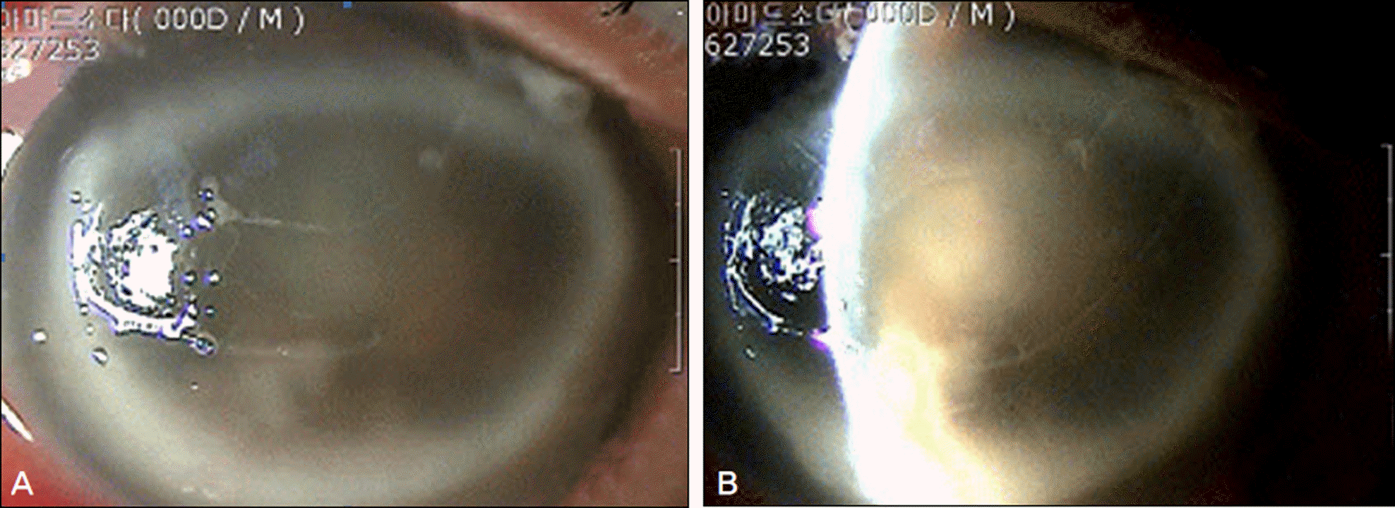
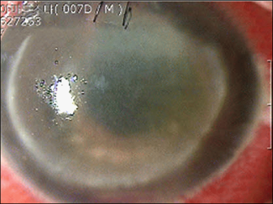
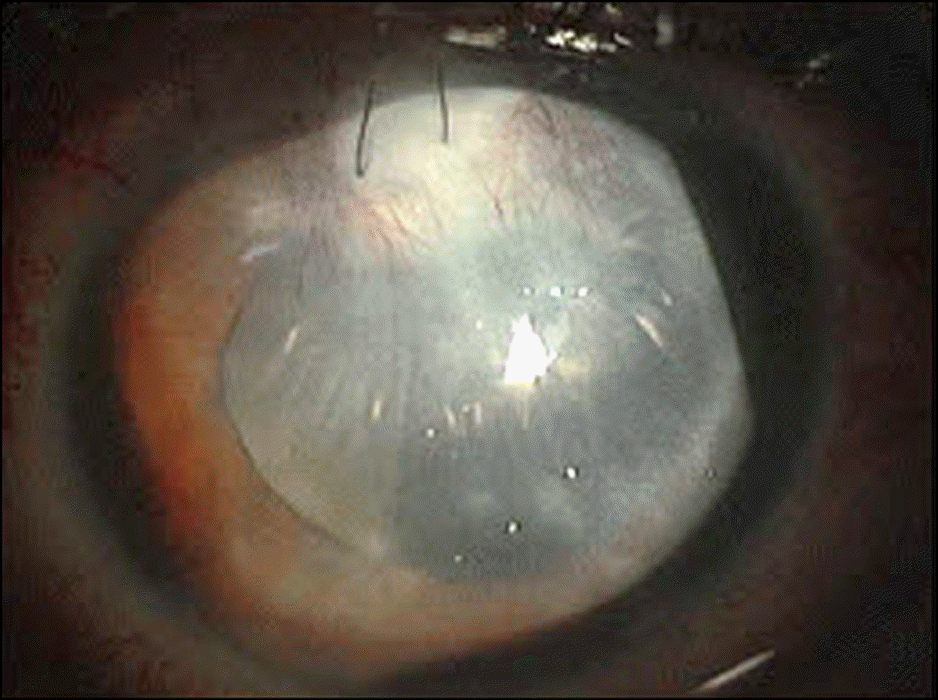
A 27-year-old man was referred for trauma caused by a fishing sinker in his right eye. On initial examination at another hospital, his visual acuity was light perception, and intraocular pressure was 50 mm Hg. On slit lamp examination, corneal edema and severe anterior chamber inflammation were observed. Consequently, the next day total pars plana vitrectomy, lensectomy, intravitreal silicone oil injection, and antibiotics injection were performed. After the operation, intraocular pressure was 15 mm Hg and the patient's pain was temporarily decreased. The presence of Shewanella algae in the vitreous culture was determined but antibiotic sensitivity was not proven. The patient received postoperative topical fortified vancomycin, ceftazidime, and tobramycin hourly and underwent intravenous antibiotic therapy. On postoperative day 25, the patient transferred to our hospital and ocular pain presented continuously. Intraocular inflammation was not severe but visual acuity was light perception because of retinal necrosis in the posterior pole. Therefore, the patient received topical fortified antibiotics and intravenous antibiotics therapy. On postoperative month 2, visual acuity was light perception and the patient's right eye progressed to pthisis bulbi.
References
1. Driebe WT Jr, Mandelbaum S, Forster RK, et al. Pseudophakic endophthalmitis. Diagnosis and management. Ophthalmology. 1986; 93:442–8.
2. Brinton GS, Topping TM, Hyndiuk RA, et al. Posttraumatic endophthalmitis. Arch Ophthalmol. 1984; 102:547–50.

3. Puliafito CA, Baker AS, Haaf J, Foster CS. Infectious endophthalmitis. Review of 36 cases. Ophthalmology. 1982; 89:921–9.

4. Holt HM, Søgaard P, Gahrn-Hansen B. Ear infection with Shewanella alga: a bacteriologic, clinical and epidemiologic study of 67 cases. Clin Microbiol Infect. 1997; 3:329–34.
5. Nozue H, Hayashi T, Hashimoto Y, et al. Isolation and characterization of Shewanella alga from human clinical specimens and emendation of the description of S.alga Simidu et al. 1990, 335. Int J Syst Bacteriol. 1992; 42:628–34.
6. Chen YS, Liu YC, Yen MY, et al. Skin and soft-tissue manifestations of Shewanella putrefaciens infection. Clin Infect Dis. 1997; 25:225–9.
7. Thompson JT, Parver LM, Enger CL, et al. Infectious endophthalmitis after penetrating injuries with retained intraocular foreign bodies. National Eye Trauma System. Ophthalmology. 1993; 100:1468–74.

8. Yoo SJ, Cho SW, Kim JW. Clinical analysis of posttraumatic endophthalmitis. J Korean Ophthalmol Soc. 2004; 45:69–78.
9. Rowsey JJ, Newsom DL, Sexton DJ, Harms WK. Endophthalmiti s: current approaches. Ophthalmology. 1982; 89:1055–66.
10. Kunimoto DY, Das T, Sharma S, et al. Microbiologic spectrum and susceptibility of isolates: part II. Posttraumatic endophthalmitis. Endophthalmitis Research Group. Am J Ophthalmol. 1999; 128:242–4.
11. Holt HM, Gahrn-Hansen B, Bruun B. Shewanella species: infections in Denmark and phenotypic characterisation. Clin Microbiol Infect. 2004; 10:suppl 3. 348–9.
12. Vogel BF, Jørgensen K, Christensen H, et al. Differentiation of Shewanella putrefaciens and Shewanella alga on the basis of whole-cell protein profiles, ribotyping, phenotypic characterization, and 16S rRNA gene sequence analysis. Appl Environ Microbiol. 1997; 63:2189–99.

13. MacDonell MT, Colwell RR. Phylogeny of the Vibrionaceae and recommendation for two new genera, Listonella and Shewanella. Syst Appl Microbiol. 1985; 6:171–82.

15. Eifrig CW, Scott IU, Flynn HW Jr, Miller D. Endophthalmitis caused by Pseudomonas aeruginosa. Ophthalmology. 2003; 110:1714–7.

16. Gram L, Bundvad A, Melchiorsen J, et al. Occurrence of Shewanella algae in Danish coastal water and effects of water temperature and culture conditions on its survival. Appl Environ Microbiol. 1999; 65:3896–900.
17. Vogel BF. A polyphasic approach in typing of Shewanella spp. and Listeria monocytogenes. PhD thesis. Royal Veterinary and Agricultural University;Lyngby, Denmark: 2000.
18. Holt HM, Gahrn-Hansen B, Bruun B. Shewanella algae and Shewanella putrefaciens: clinical and microbiological characteristics. Clin Microbiol Infect. 2005; 11:347–52.

19. Khashe S, Janda JM. Biochemical and pathogenic properties of Shewanella alga and Shewanella putrefaciens. J Clin Microbiol. 1998; 36:783–7.
20. Weyant RS, Moss CW, Weaver RE, et al. Identification of unusual pathogenic gram-negative aerobic and facultatively anaerobic bacteria. Baltimore: Williams & Wilkins;1996.
Figure 1.
Photograph at the first visit. (A) Corneal ring infiltration and anterior chamber cells and flare were seen in the right eye. (B) Corneal edema was seen and inflammatory material was filled in the anterior chamber of the right eye.


 PDF
PDF ePub
ePub Citation
 Print
Print


 XML Download