

 PDF
PDF ePub
ePub Citation
 Print
Print


Abstract
Purpose
The effects of AmniSite-Lens on wound healing were evaluated for a burn wound on a rabbit cornea.
Methods
A chemical burn was inflicted on the cornea of rabbits using 0.1N NaOH and a superficial keratectomy with trephine was performed. The control group consisted of rabbits with a bandage contact lens (Focus Lens) after the operation. In the other group, the AmniSite-Le was applied on the rabbits' cornea. The rabbits were evaluated for the following: 1) the time of epithelialization; 2) the grade of corneal opacity; and 3) the histological analysis by evaluation of inflammatory reaction and apoptotic keratocytes.
Results
In the alkali-burn model, the time of epithelialization in the AmniSite-Lens group was not statistically significant compared with the bandage contact lens group. There was no difference of corneal opacity at postoperative week 1. The corneal opacity in the AmniSite-Lens group was clearer than the bandage contact lens group at postoperative weeks 4 and 8 and the difference of corneal opacity was statistically significant. In the keratectomy model, the time of epithelialization in the AmniSite-Lens group was not statistically significant compared with the bandage contact lens group. The corneal opacity in the AmniSite-Lens group was clearer than the bandage contact lens group at postoperative weeks 1 and 4 and the difference of corneal opacity was statistically significant.
Go to : 
References
1. Shimmura S, Shimazaki J, Ohashi Y, Tsubota K. Antiinflammatory effects of amniotic membrane transplantation in ocular surface disorders. Cornea. 2001; 20:408–13.

2. Kim JS, Kim JC, Na BK, et al. Amniotic membrane patching promotes healing and inhibits proteinase activity on wound healing following acute corneal alkali burn. Exp Eye Res. 2000; 70:329–37.
3. Gris O, del Campo Z, Wolley-Dod C, et al. Amniotic membrane implantation as a therapeutic contact lens for the treatment of epithelial disorders. Cornea. 2002; 21:22–7.
4. Takano Y, Fukagawa K, Miyake-Kashima M, et al. Dramatic healing of an allergic corneal ulcer persistent for 6 months by amniotic membrane patching in a patient with atopic keratoconjunctivitis: a case report. Cornea. 2004; 23:723–5.
5. Choi YS, Kim JY, Wee WR, Lee JH. Effect of the application of human amniotic membrane on rabbit corneal wound healing after excimer laser photorefractive keratectomy. Cornea. 1998; 17:389–95.
6. Woo HM, Kim MS, Kweon OK, et al. Effects of amniotic membrane on epithelial wound healing and stromal remodelling after excimer laser keratectomy in rabbit cornea. Br J Ophthalmol. 2001; 85:345–9.
7. Seo JW, Ko BW, Lee DJ, Park WC. The effects of amniotic membrane contact lens for cornea wound healing. J Korean Ophthalmol Soc. 2009; 50:989–95.
8. De Rotth A. Plastic repair of conjunctival defects with fetal membranes. Arch Ophthalmol. 1940; 23:525–5.
9. Batlle JF, Perdomo FJ. Placental membranes as a conjunctival substitute. Ophthalmology. 1993; 100:107.
10. Kim JC, Tseng SC. Transplantation of preserved human amniotic membrane for surface reconstruction in severely damaged rabbit corneas. Cornea. 1995; 14:473–84.
11. Nakamura T, Inatomi T, Sekiyama E, et al. Novel clinical application of sterilized, freeze-dried amniotic membrane to treat patients with pterygium. Acta Ophthalmol Scand. 2006; 84:401–5.
12. Park M, Kim S, Kim IS, Son D. Healing of a porcine burn wound dressed with human and bovine amniotic membranes. Wound Repair Regen. 2008; 16:520–8.
Go to :
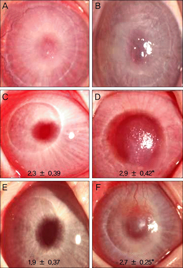 | Figure 2.Photographs showing corneal opacity at 1 day (A, B), 1 week (C, D), and 4 weeks (E, F) after superficial keratectomy. Corneal opacity was improved in all groups, but in the bandage contact lens group (B, D, F), the opacity was more prominent than in AmniSite-Lens group (A, C, E). The difference of cornea opacity was statistically significant at 1 week and 4 weeks (*p < 0.05). |
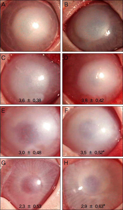 | Figure 3.Photographs showing corneal opacity at 1 day (A, B), 1 week (C, D), 4 weeks (E, F), and 8 weeks (G, H) after alkali burn. Corneal opacity was improved in all groups, but in the bandage contact lens group (B, D, G, H), the opacity was more prominent than in AmniSite-Lens group (A, C, E, G). The difference of cornea opacity was statistically significant at 4, 8 weeks (*p < 0.05). |
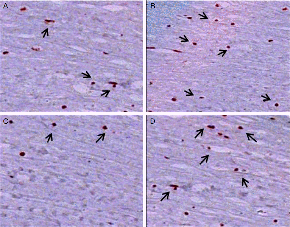
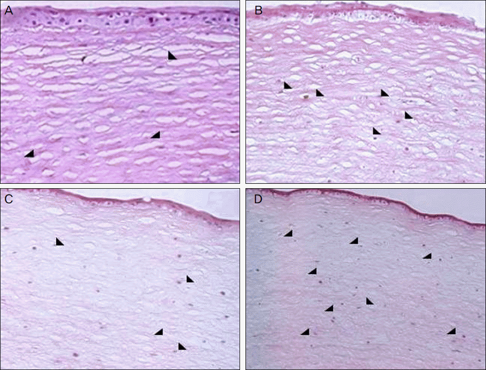 | Figure 4.Histopathologic findings of rabbit cornea with hematoxylin and eosin staining (×200). M ild infiltration of inflammatory cells was detected in the AmniSite-Lens (A) and bandage contact lens group (B) of the alkali-burn model and the AmniSite-Lens (C) and bandage contact lens group (D) of the superficial keratectomy model. |
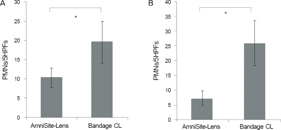 | Figure 5.Comparison of PMNs infiltration in the stroma. There was significant difference between the AmniSite-Lens and bandage CL group (*p < 0.05). (A) Alkali burn model, (B) Superficial keratectomy model. PM Ns = polymorphonuclear cells; HPF = high power field. |
 | Figure 6.Histopathologic findings of the rabbit cornea with TUNEL stain (×400). The lesser TUNEL positive cells were detected in the AmniSite-Lens (A) than in the bandage contact lens group (B) of the alkali-burn model. Also, lesser TUNEL positive cells were detected in the AmniSite-Lens (C) than in the bandage contact lens group (D) of the superficial keratectomy model. |
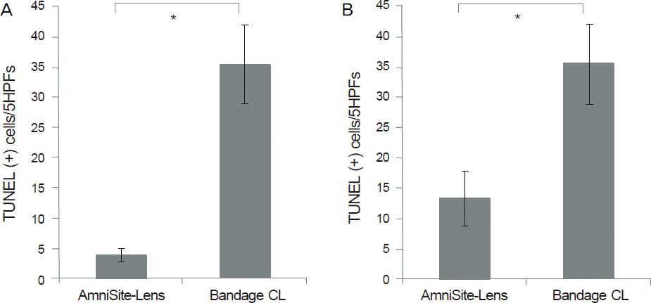 | Figure 7.Comparison of TUNEL positive cell counts. More TUNEL positive cells were seen in the bandage contact lens group. There was significant difference between the AmniSite-Lens and the bandage CL group (*p < 0.05). (A) Alkali burn model, (B) Superficial keratectomy model. HPF = high power field. |
 XML Download