Abstract
Purpose
The changes in anterior chamber depth (ACD) and refractive error in pseudophakia with 6 types of intraocular lenses (IOLs) after cataract surgery were compared in the present study.
Methods
The medical records of 108 eyes (73 patients) who underwent cataract surgery with 6 types of IOLs, 5 types of single-piece IOLs and 1 type of 3-piece IOLs between March 2007 and April 2010 were retrospectively reviewed. ACD and refractive error were measured preoperatively, and at 1 day, 1 week, 1 month and 3 months postoperatively and the data were extracted and analyzed.
Results
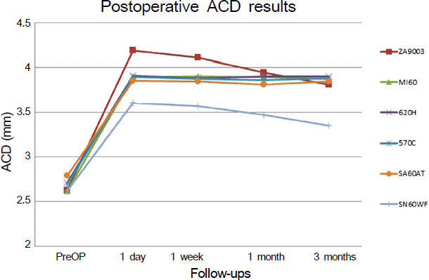
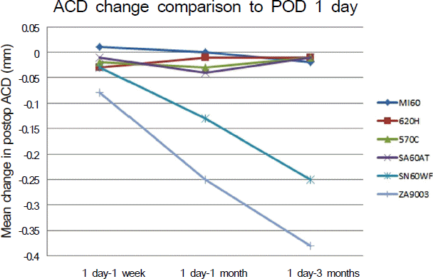
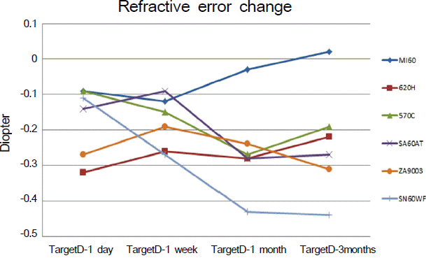
In the case of the SN60WF lens, the ACD was significantly shallow as compared to other IOLs at each follow-up period and refractive error was significantly myopic at 3 months postoperatively. In the case of SN60WF and ZA9003 lenses, the ACD was significantly changed at 3 months postoperatively from 1 day postoperatively. In the case of the MI60 lens, refractive error was slightly hyperopic, but not statistically significant.
Go to : 
References
1. John T, Shah AA. New surgical technique: upside-down phacoemulsification with posterior chamber intraocular lens and Descemet's stripping automated endothelial keratoplasty. Ann Ophthalmol (Skokie). 2009; 41:16–23.
2. Olsen T, Thim K, Corydon L. Theoretical versus SRK I and SRK II calculation of intraocular lens power. J Cataract Refract Surg. 1990; 16:217–25.

3. Remsch H, Kampmeier J, Muche R, et al. [Comparison of the optical coherence method (Zeiss IOL-Master) with two ultrasono-graphic biometric methods for the calculation of posterior chamber intraocular lenses after phacoemulsification as part of clinical routine]. Klin Monbl Augenheilkd. 2004; 221:837–42.
4. Saka N, Moriyama M, Shimada N, et al. Changes of axial length measured by IOL master during 2 years in eyes of adults with pathologic myopia. Graefes Arch Clin Exp Ophthalmol. 2012.
5. van der Linden JW, van Velthoven M, van der Meulen I, et al. Comparison of a new-generation sectorial addition multifocal intraocular lens and a diffractive apodized multifocal intraocular lens. J Cataract Refract Surg. 2012; 38:68–73.

6. López-Gil N, Montés-Micó R. New intraocular lens for achromatizing the human eye. J Cataract Refract Surg. 2007; 33:1296–302.

7. Holladay JT, Moran JR, Kezirian GM. Analysis of aggregate surgically induced refractive change, prediction error, and intraocular astigmatism. J Cataract Refract Surg. 2001; 27:61–79.

8. Chen MJ, Liu YT, Tsai CC, et al. Relationship between central cor-neal thickness, refractive error, corneal curvature, anterior chamber depth and axial length. J Chin Med Assoc. 2009; 72:133–7.

9. Olsen T. Prediction of the effective postoperative (intraocular lens) anterior chamber depth. J Cataract Refract Surg. 2006; 32:419–24.

10. Watson A, Armstrong R. Contact or immersion technique for axial length measurement? Aust N Z J Ophthalmol. 1999; 27:49–51.
11. Høvding G, Natvik C, Sletteberg O. The refractive error after implantation of a posterior chamber intraocular lens. The accuracy of IOL power calculation in a hospital practice. Acta Ophthalmol. 1994; 72:612–6.

12. Tanaka T. Refractive error in VA-60 BB acrylic intraocular lenses. J Cataract Refract Surg. 2005; 31:1673–4.
13. Erickson P. Effects of intraocular lens position errors on postoperative refractive error. J Cataract Refract Surg. 1990; 16:305–11.

14. Oh SH, Kim JK, Lee DH. The clinical results of hydrophobic single-piece acrylic intraocluar lenses after cataract surgery. J Korean Ophthalmol Soc. 2004; 45:2007–13.
15. Chae JK, Jang JW, Choi TH, Lee HB. Changes in refraction and anterior chamber depth according to the type of intraocular lenses. J Korean Ophthalmol Soc. 2006; 47:1935–42.
16. Moon SH, Lee DH, Lew HM. The change of anterior chamber depth according to the types of intraocular lens. J Korean Ophthalmol Soc. 1998; 39:2280–5.
17. Rose LT, Moshegov CN. Comparison of the Zeiss IOLMaster and applanation A-scan ultrasound: biometry for intraocular lens calculation. Clin Experiment Ophthalmol. 2003; 31:121–4.

18. Brandser R, Haaskjold E, Drolsum L. Accuracy of IOL calculation in cataract surgery. Acta Ophthalmol Scand. 1997; 75:162–5.

19. Szaflik J, Kaminska A, Gajda S, Jedruch A. [Accuracy of the SRK II, SRK/T, Holladay and Hoffer Q IOL power calculation formulas -in hyperopic patients after phacoemulsification]. Klin Oczna. 2005; 107:615–9.
20. Zaldivar R, Shultz MC, Davidorf JM, Holladay JT. Intraocular lens power calculations in patients with extreme myopia. J Cataract Refract Surg. 2000; 26:668–74.

21. Armstrong TA. Refractive effect of capsular bag lens placement with the capsulorhexis technique. J Cataract Refract Surg. 1992; 18:121–4.

22. Minassian DC, Rosen P, Dart JK, et al. Extracapsular cataract extraction compared with small incision surgery by phacoemulsification: a randomised trial. Br J Ophthalmol. 2001; 85:822–9.

23. Shammas HJ, Shammas MC, Garabet A, et al. Correcting the cor-neal power measurements for intraocular lens power calculations after myopic laser in situ keratomileusis. Am J Ophthalmol. 2003; 136:426–32.

24. Celikkol L, Ahn D, Celikkol G, Feldman ST. Calculating intraocular lens power in eyes with keratoconus using videokeratography. J Cataract Refract Surg. 1996; 22:497–500.

25. Shioya M, Ogino N, Shinjo U. Change in postoperative refractive error when vitrectomy is added to intraocular lens implantation. J Cataract Refract Surg. 1997; 23:1217–20.

26. Holladay JT, Prager TC, Ruiz RS, et al. Improving the predictability of intraocular lens power calculations. Arch Ophthalmol. 1986; 104:539–41.

27. Whitehouse G. Effect of lens style on postoperative refractive astigmatism after small incision cataract surgery. Clin Experiment Ophthalmol. 2000; 28:290–2.

28. Petternel V, Menapace R, Findl O, et al. Effect of optic edge design and haptic angulation on postoperative intraocular lens position change. J Cataract Refract Surg. 2004; 30:52–7.

29. Olsen T, Corydon L, Gimbel H. Intraocular lens power calculation with an improved anterior chamber depth prediction algorithm. J Cataract Refract Surg. 1995; 21:313–9.

30. Olsen T. Sources of error in intraocular lens power calculation. J Cataract Refract Surg. 1992; 18:125–9.

31. Arai M, Ohzuno I, Zako M. Anterior chamber depth after posterior chamber intraocular lens implantation. Acta Ophthalmol. 1994; 72:694–7.

32. Wirtitsch MG, Findl O, Menapace R, et al. Effect of haptic design on change in axial lens position after cataract surgery. J Cataract Refract Surg. 2004; 30:45–51.

33. Landers J, Liu H. Choice of intraocular lens may not affect refractive stability following cataract surgery. Clin Experiment Ophthalmol. 2005; 33:34–40.
34. Lee JY, Lee SH, Chung SK. Decentration, tilt and anterior chamber depth: aspheric vs spheric acrylic intraocular lens. J Korean Ophthalmol Soc. 2009; 50:852–7.

35. Amzallag T, Pynson J. [Lens biomaterials for cataract surgery]. J Fr Ophtalmol. 2007; 30:757–67.
Go to : 
 | Figure 1.The postoperative mean anterior chamber depth (ACD) in 6 types of IOL. In the SN60W F, the ACD is significantly shallow as compared to other IOLs at each follow-up period. |
 | Figure 2.The postoperative differences of mean anterior chamber depth after cataract surgery. In the SN60W F and ZA9003, there is significant change in ACD at 3 months postoperatively from 1 day postoperatively. |
 | Figure 3.The postoperative refractive error (mean spherical equivalent differences) between the target refraction and the actual refraction at each period. In the SN60W F, refractive error is significantly myopic at 3 months postoperativlely. In the MI60, refractive error is slightly hyperopic, but not statistically significant. |
Table 1.
Preoperative characteristics of 6 types of IOL
| Preoperative Characteristics | Types of IOL | ||||||
|---|---|---|---|---|---|---|---|
| SA60AT | SN60WF | MI60 | 620H | 570C | ZA9003 | p-value | |
| Eyes/N | 29/18 | 18/14 | 6/3 | 10/6 | 14/8 | 31/24 | |
| Sex (M/F) | 10:19 | 8:10 | 4:02 | 6:04 | 8:06 | 18:13 | 0.40∗ |
| Age (year) | 68.26 ± 7.23 | 62.44 ± 8.50 | 80.00 ± 11.23 | 67.20 ± 9.41 | 71.57 ± 10.27 | 69.13 ± 8.57 | 0.13† |
| AxL (mm) | 22.87 ± 0.67 | 22.94 ± 0.53 | 22.89 ± 0.62 | 23.04 ± 0.57 | 23.08 ± 0.49 | 23.09 ± 0.61 | 0.36† |
| ACD (mm) | 2.79 ± 0.45 | 2.62 ± 0.37 | 2.62 ± 0.46 | 2.71 ± 0.47 | 2.69 ± 0.39 | 2.63 ± 0.46 | 0.24† |


 PDF
PDF ePub
ePub Citation
 Print
Print


 XML Download