

 PDF
PDF ePub
ePub Citation
 Print
Print


Abstract
Purpose
To analyze the morphology and density of corneal tissue in patients with bullous keratopathy using in vivo confocal microscopy (IVCM).
Case summary
Four eyes of 4 patients with clinically diagnosed bullous keratopathy were examined using IVCM. Cross-sectioned corneal images of the corneal epithelium, Bowman’s layer, stromal layer, Descemet’s membrane, and endothelium were evaluated. In bullous keratopathy patients, the outline of corneal epithelial wing cells was blurred. Several hyper-reflective whitish dots were found at the corneal epithelial level and a reduction in the density and thickness of the corneal nerves was observed. There was also a reduction in the density of the stromal keratocytes. The outline of the stromal keratocytes was blurred. Some keratocytes resembled myofibroblast. The hypo-reflective vertical strands were observed in the stroma adjacent to the Descemet’s membrane. Endothelium showed hyper-reflectivity with associated increased endothelial pleomorphism and polymegathism.
Conclusions
In patients with bullous keratopathy, IVCM showed tissue edema in the corneal epithelium, stroma, and endothelium. Hyper-reflectivity appearances were noted due to the increase in the metabolic rate of the cells or ex-tracellular matrix accumulation. Corneal endothelium showed morphological changes.
Go to : 
References
1. Shimazaki J, Amano S, Uno T. . National survey on bullous keratopathy in Japan. Cornea. 2007; 26:274–8.

2. Gonçalves ED, Campos M, Paris F. . Bullous keratopathy: etio-pathogenesis and treatment. Arq Bras Oftalmol. 2008; 71:61–4.
3. Waring GO 3rd, Rodrigues MM. Patterns of pathologic response in the cornea. Surv Ophthalmol. 1987; 31:262–6.
4. Choi SH, Lee YW, Kim HM. . Epidemiologic studies of kera-toplasty in Korea. J Korean Ophthalmol Soc. 2006; 47:538–47.
5. Yuen HKL, Rassier CE, Jardeleza MSR. . A morphologic study of Fuchs dystrophy and bullous keratopathy. Cornea. 2005; 24:319–27.
6. Al-Aqaba M, Alomar T, Lowe J, Dua HS. Corneal nerve aberra-tions in bullous keratopathy. Am J Ophthalmol. 2011; 151:840–9.e1.
7. Borboli S, Colby K. Mechanisms of disease: Fuchs' endothelial dystrophy. Ophthalmol Clin North Am. 2002; 15:17–25.
8. Bergmanson JP, Sheldon TM, Goosey JD. Fuchs' endothelial dys-trophy: a fresh look at an aging disease. Ophthalmic Physiol Opt. 1999; 19:210–22.
9. Senoo T, Takahashi K, Chiba K. . Stimulation of corneal endo-thelial cell proliferation by interleukins, complete mitogens and corneal parenchymal cell-derived factors. Nippon Ganka Gakkai Zasshi. 1996; 100:845–52.
10. Fini ME. Keratocyte and fibroblast phenotypes in the repairing cornea. Prog Retin Eye Res. 1999; 18:529–51.
11. Thalmann-Goetsch A, Engelmann K, Bednarz J. Comparative study on the effects of different growth factors on migration of bovine corneal endothelial cells during wound healing. Acta Ophthalmol Scand. 1997; 75:490–5.
12. Imanishi J, Kamiyama K, Iguchi I. . Growth factors: importance in wound healing and maintenance of transparency of the cornea. Prog Retin Eye Res. 2000; 19:113–29.
13. Ljubimov AV, Burgeson RE, Butkowski RJ. . Extracellular matrix alterations in human corneas with bullous keratopathy. Invest Ophthalmol Vis Sci. 1996; 37:997–1007.
14. Waring GO 3rd, Rodrigues MM, Laibson PR. Corneal dystrophies. II. Endothelial dystrophies. Surv Ophthalmol. 1978; 23:147–68.
15. Morishige N, Yamada N, Zhang X. . Abnormalities of stromal structure in the bullous keratopathy cornea identified by second harmonic generation imaging microscopy. Invest Ophthalmol Vis Sci. 2012; 53:4998–5003.
16. Yagmur M, Okay O, Sizmaz S. . In vivo confocal microscopy: corneal changes of hydrogel contact lens wearers. Int Ophthalmol. 2011; 31:377–83.
17. Morishige N, Takahashi N, Nobuhiko C, Nishida T. Quantitative evaluation of corneal epithelial oedema by confocal microscopy. Clin Exper Ophthalmol. 2009; 37:249–53.
Go to :
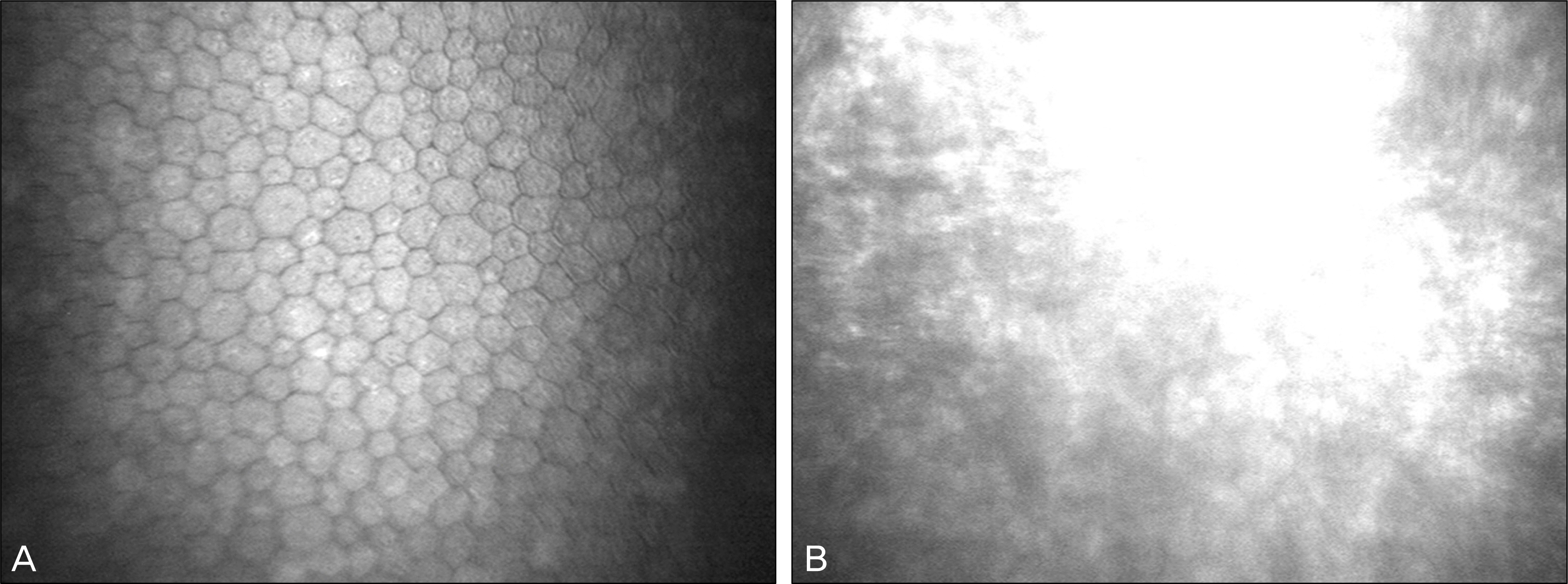
 | Figure 1.
In vivo confocal biomicroscopy of the corneal epithelium. (A) Normal appearance of the corneal epithelial wing cells seen in a healthy control. (B) The outline of corneal epithelial wing cells is blurred. Diffuse corneal edema is noted. Some hyper-reflective whitish dots are found. |
 | Figure 2.
In vivo confocal biomicroscopy of the corneal nerves. (A) Normal appearance of the sub-basal nerve plexus seen in a healthy control. (B) Sub-basal nerve plexus appearance in bullous keratopathy. There is a reduction in the density and thickness of the nerves. The outline of corneal keratocyte is blurred. |
 | Figure 3.
In vivo confocal biomicroscopy of the corneal stroma. (A) Normal appearance of the stroma and keratocytes seen in a healthy control. (B) There is a reduction in the density of the keratocytes in the anterior stroma. The outline of the stromal keratocytes is blurred. Some keratocytes resemble myofibroblast.(C) The hypo-reflective vertical strands are seen in the stroma adjacent to the descemet membrane. |
 XML Download