

 PDF
PDF ePub
ePub Citation
 Print
Print


Abstract
Purpose
To describe the rate of surgical intervention to the intranasal structures for making a sufficient bony ostium in endonasal dacryocystorhinostomy.
Methods
Video records of 52 Korean patients (66 eyes) who underwent endonasal dacryocystorhinostomy between January 2010 and December 2010 for primary nasolacrimal duct obstruction were reviewed. To locate the internal common punctum in the lateral nasal wall, a transcanalicular illumination device consisting of disposable vitrectomy light pipe was introduced horizontally through the canaliculus. The relative position of the internal common punctum to the operculum of the middle turbinate was divided into 4 types and analyzed according to the intranasal surgical procedures necessary.
Results
Internal common punctum was located posterior-superior to the operculum of the middle turbinate in 37 cases (56%), anterior-superior in 16 cases (24%), posterior-inferior in 11 cases (17%) and anterior-inferior in 2 cases (3%). To expose the bony ostium, partial removal of the operculum of the middle turbinate was required in 63 cases (95%), anterior middle turbinectomy in 43 cases (65%), uncinectomy in 62 cases (94%) and opening of the agger nasi cell in 45 cases (68%).
Conclusions
In a majority of patients, partial removal of the middle turbinate, uncinate process and agger nasi cell were necessary to create a sufficient bony ostium. The transcanalicular illumination device is useful to locate the lacrimal sac and can be helpful in understanding the intranasal structures which need to be removed during surgery.
Go to : 
References
1. Detorakis ET, Drakonaki EE, Bizakis I, et al. MRI evaluation of lacrimal drainage after external and endonasal dacryocystorhinostomy. Ophthal Plast Reconstr Surg. 2009; 25:289–92.

2. You IC, Jeong SK, Seo JJ.Anatomical study of lacrimal passage using computed tomography. J Korean Ophthalmol Soc. 2002; 43:2112–8.
3. Woo KI, Maeng HS, Kim YD.Characteristics of intranasal structures for endonasal dacryocystorhinostomy in asians. Am J Ophthalmol. 2011; (152):491–8.
4. Moore WM, Bentley CR, Olver JM.Functional and anatomic results after two types of endoscopic endonasal dacryocystorhinostomy: surgical and holmium laser. Ophthalmology. 2002; (109):1575–82.
5. Fayet B, Racy E. Assouline M. Systematic unciformectomy for a standardized endonasal dacryocystorhinostomy. Ophthalmology. 2002; (109):530–6.
6. Fayet B, Racy E, Assouline M, Zerbib M.Surgical anatomy of the lacrimal fossa. A prospective computed tomodensitometry scan analysis. Ophthalmology. 2005; (112):1119–28.
7. Metson R.Endoscopic surgery for lacrimal obstruction. Otolaryngol Head Neck Surg. 1991; (104):473–9.
8. Metson R, Woog JJ, Puliafito CA.Endoscopic laser dacryocystorhinostomy. Laryngoscope. 1994; 104((3 Pt 1)):269–74.
9. Mickelson SA, Kim DK, Stein IM.Endoscopic laser-assisted dacryocystorhinostomy. Am J Otolaryngol. 1997; 18:107–11.
10. Gustafson RO, Bartley GB.A plea for preservation of the middle turbinate during dacryocystorhinostomy. Ophthal Plast Reconstr Surg. 1999; 15:75–6.
11. Tsirbas A, Wormald PJ.Endonasal dacryocystorhinostomy with mucosal flaps. Am J Ophthalmol. 2003; 135:76–83.
12. Cokkeser Y, Evereklioglu C, Er H.Comparative external versus endoscopic dacryocystorhinostomy: results in 115 patients (130 eyes). Otolaryngol Head Neck Surg. 2000; 123:488–91.
13. Benger R, Forer M.Endonasal dacryocystorhinostomy–primary and secondary. Aust N Z J Ophthalmol. 1993; 21:157–9.
14. Tsirbas A, Wormald PJ.Mechanical endonasal dacryocystorhinostomy with mucosal flaps. Br J Ophthalmol. 2003; 87:43–7.
15. Tsirbas A, Davis G, Wormald PJ.Mechanical endonasal dacryocystorhinostomy versus external dacryocystorhinostomy. Ophthal Plast Reconstr Surg. 2004; 20:50–6.
16. Wanamaker HH.Role of Haller's cell in headache and sinus disease: a case report. Otolaryngol Head Neck Surg. 1996; 114:324–7.
17. Yung MW, Hardman-Lea S.Endoscopic inferior dacryocystorhinostomy. Clin Otolaryngol Allied Sci. 1998; 23:152–7.
18. Fayet B, Racy E.[Is the uncinate process resection the key to endo-nasal dacryocystorhinostomy?]. J Fr Ophtalmol. 2000; 23:433–6.
19. Fayet B, Racy E, Assouline M.Complications of standardized endonasal dacryocystorhinostomy with unciformectomy. Ophthalmology. 2004; 111:837–45.
20. Kim JL, Yang JW.Clinical consideration of uncinectomy for endo-nasal dacryocystorhinostomy. J Korean Ophthalmol Soc. 2008; 49:871–7.
21. Yang JW, Oh HN.Success rate and complications of endonasal dacryocystorhinostomy with unciformectomy. Graefes Arch Clin Exp Ophthalmol. 2012; 250:1509–13.
22. Anon JB, Rontal M, Zinreich SJ.Anatomy of the paranasal sinuses. Thieme. 1996; 29–34.
23. Kennedy DW, Bolger WE, Zinreich SJ.Diseases of the sinuses: diagnosis and management. Hamilton, Ontario, Canada: BC Decker;2001. –17. p. 24
24. Levine H, Clemente M.Sinus surgery: endoscopic and microscopic approaches. Stuttgart: Thieme. 2005; 312–5.
25. Rebeiz EE, Shapshay SM, Bowlds JH, Pankratov MM.Anatomic guidelines for dacryocystorhinostomy. Laryngoscope. 1992; 102:1181–4.
26. Wormald PJ, Kew J, Van Hasselt A.Intranasal anatomy of the nasolacrimal sac in endoscopic dacryocystorhinostomy. Otolaryngol Head Neck Surg. 2000; 123:307–10.
27. Rebeiz EE, Shapshay SM, Bowlds JH, Pankratov MM.Anatomic guidelines for dacryocystorhinostomy. Laryngoscope. 1992; 102:1181–4.
28. Sprekelsen MB, Barberán MT.Endoscopic dacryocystorhinostomy: surgical technique and results. Laryngoscope. 1996; 106((2 Pt 1)):187–9.
29. Cunningham MJ, Woog JJ.Endonasal endoscopic dacryocystorhinostomy in children. Arch Otolaryngol Head Neck Surg. 1998; 124:328–33.
30. Fayet B, Vericel R, Racy E, et al. DCR par voie endonasale couple´e a` la transillumination canaliculaire. Bull Soc Ophtalmol Fr. 1998; 98:223–6.
31. Whittet HB, Shun-Shin GA, Awdry P.Functional endoscopic transnasal dacryocystorhinostomy. Eye. 1993; 7((Pt 4)):545–9.
32. Boush GA, Lemke BN, Dortzbach RK.Results of endonasal laser-assisted dacryocystorhinostomy. Ophthalmology. 1994; 101:955–9.
33. Rebeiz EE, Shapshay SM, Bowlds JH, Pankratov MM.Anatomic guidelines for dacryocystorhinostomy. Laryngoscope. 1992; 102:1181–4.
34. Gonnering RS, Lyon DB, Fisher JC.Endoscopic laser-assisted lacrimal surgery. Am J Ophthalmol. 1991; 111:152–7.
35. Lee JJ, Woo KI, Kim YD.Middle turbinectomy during dacryocystorhinostomy. J Korean Ophthalmol Soc. 1997; 38:710–4.
36. Kwon S, Baek SH.Clinical evaluation of endoscopic endonasal dacryocystorhinostomy. J Korean Ophthalmol Soc. 2004; 45:1403–8.
37. Chung ER, Lee KT, Choi WC.Success rate of endonasal dacryocystorhinostomy based on the location of the lacrimal sac. J Korean Ophthalmol Soc. 2002; 43:2000–4.
38. Ercan I, Cakir BO, Sayin I, et al. Relationship between the superior attachment type of uncinate process and presence of agger nasi cell: a computer-assisted anatomic study. Otolaryngol Head Neck Surg. 2006; 134:1010–4.
39. Shin HM, Lew H, Yun YS.Surgical result of endoscopic dacryocystorhinostomy according to opening size of nasal mucosa. J Korean Ophthalmol Soc. 2006; 47:175–80.
Go to :
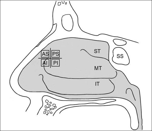 | Figure 1.Comparative position of the internal common punctum divided by 4 quadrants. AS = anterior-superior; AI = anterior-inferior; PS = posterior-superior; PI = posterior-inferior; ST = superior turbinate; MT = middle turbinate; IT = inferior turbinate; SS = sphenoidal sinus. |
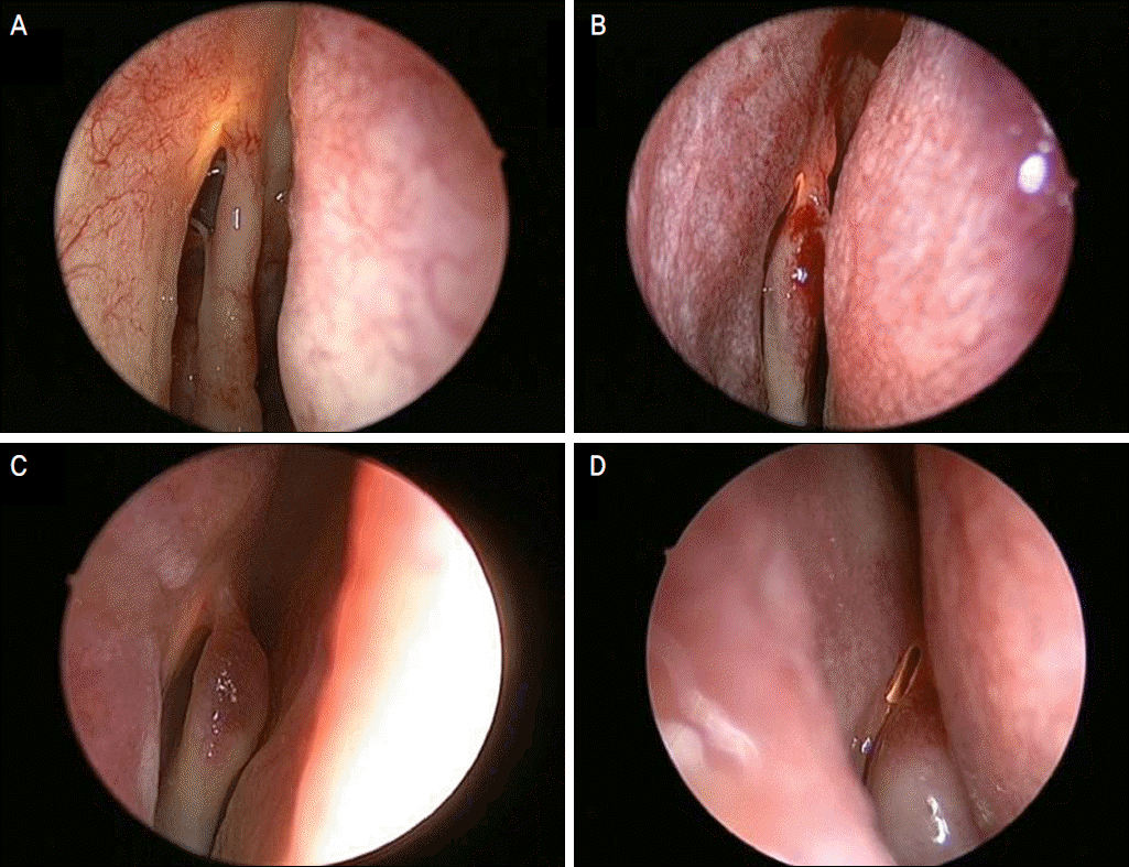 | Figure 2.Relative position of the internal common punctum to the operculum of the middle turbinate. (A) Anterior-superior, (B) Posterior-superior, (C) Anterior-inferior, (D) Posterior-inferior position. |
Table 1.
The rates of intranasal procedures according to the relative positions of the internal common punctum to the operculum of the middle turbinate
 XML Download