Abstract
Purpose
To evaluate the efficacy, safety, and satisfaction of patients who underwent LASIK for presbyopia correction in myopic patients using aspheric micro-monovision.
Methods
LASIK for presbyopic correction using aspheric micro-monovision was performed in 18 patients between December 2010 and December 2011. Distance, intermediate, and near visual acuity, refractive change, and patient’s satisfaction were evaluated for at least 12 months after the surgery.
Results
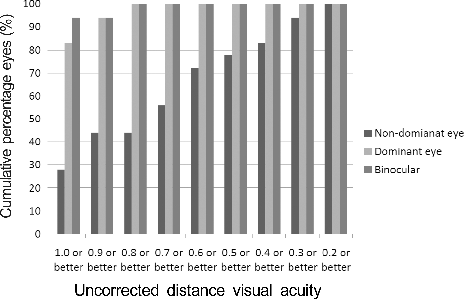
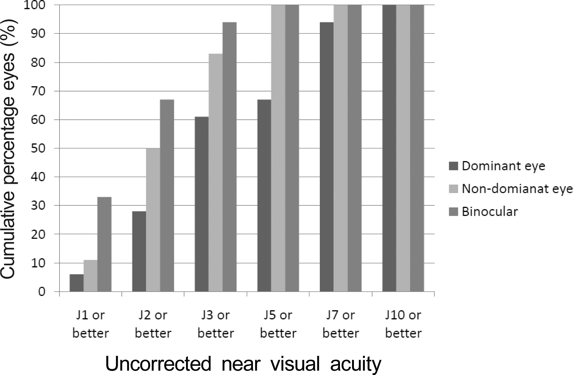
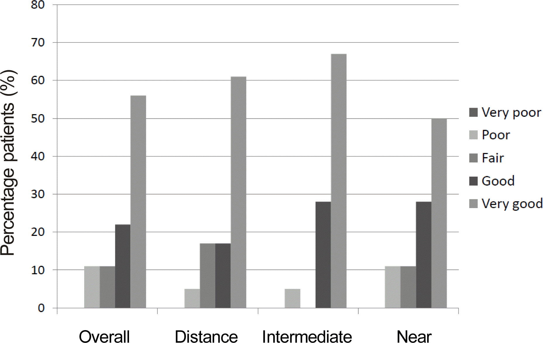
Among dominant eyes, 100% achieved uncorrected distance and intermediate visual acuity of 0.8 or better and 100% of the eyes achieved 0.8 or better binocularly. In the non-dominant eyes, 83% achieved uncorrected near visual acuity of J3 or better, and 94% of the eyes achieved J3 or better binocularly. Postoperatively, the mean manifest refraction spherical equivalent (MRSE) of the dominant eyes were -0.09 ± 0.35D, -0.17 ± 0.42D, and -0.17 ± 0.47D at 1, 6 and 12 months, respectively. The MRSE of the non-dominant eyes were -0.94 ± 0.53D, -1.03 ± 0.56D, and -1.02 ± 0.50D at post-operative 1, 6, and 12 months, respectively, without significant regression. After surgery, the patient’s overall satisfaction score was good (4.2 out of 5).
References
1. Kim MG, Kim TJ, Park YG, Lee YS. Presbyopic Contact Lens Fitting. Lee YS, editor. Contact lens: Principles and practice. 1st ed.Seoul: Naewae haksool;2007. p. 133–41.
3. Lee HY, Her J. Clinical evaluation of monovision after cataract surgery. J Korean Ophthalmol Soc. 2008; 49:1437–42.

4. Waring GO 4th. Correction of presbyopia with a small aperture corneal inlay. J Refract Surg. 2011; 27:842–5.

5. Alió JL, Chaubard JJ, Caliz A. . Correction of presbyopia by technovision central multifocal LASIK (presby-LASIK). J Refract Surg. 2006; 22:453–60.

6. Harris MG, Classe JG. Clinicolegal considerations of monovision. J Am Optom Assoc. 1988; 59:491–5.
7. Stahl JE. Conductive keratoplasty for presbyopia: 3-year results. J Refract Surg. 2007; 23:905–10.

8. Beddow DR, Martin SJ, Pfeiffer CH. Presbyopic patients and single vision contact lenses. South J Optom. 1966; 8:9–11.
10. Miranda D, Krueger RR. Monovision laser in situ keratomileusis for pre-presbyopic and presbyopic patients. J Refract Surg. 2004; 20:325–8.

11. Reinstein DZ, Archer TJ, Gobbe M. LASIK for myopic astigma-tism and presbyopia using non-linear aspheric micro-monovision with the Carl Zeiss Meditec MEL 80 Platform. J Refract Surg. 2011; 27:23–37.

12. Reinstein DZ, Couch DG, Archer TJ. LASIK for hyperopic astig-matism and presbyopia using micro-monovision with the Carl Zeiss Meditec MEL80 platform. J Refract Surg. 2009; 25:37–58.

13. Reinstein DZ, Carp GI, Archer TJ, Gobbe M. LASIK for presbyopia correction in emmetropic patients using aspheric ablation profiles and a micro-monovision protocol with the carl zeiss meditec MEL 80 and visumax. J Refract Surg. 2012; 28:531–9.

14. Wright KW, Guemes A, Kapadia MS, Wilson SE. Binocular func-tion and patient satisfactionafter monovision induced by myopic photorefractive keratectomy. J Cataract Refract Surg. 1999; 25:177–82.

15. Goldberg DB. Comparison of myopes and hyperopes after laser in situ keratomileusis monovision. J Cataract Refract Surg. 2003; 29:1695–701.

16. Reilly CD, Lee WB, Alvarenga L. . Surgical monovision and monovision reversal in LASIK. Cornea. 2006; 25:136–8.

18. Yasuda A, Yamaguchi T. Steepening of corneal curvature with contraction of the ciliary muscle. J Cataract Refract Surg. 2005; 31:1177–81.

19. Glasser A, Troilo D, Howland HC. The mechanism of corneal ac-commodation in chicks. Vision Res. 1994; 34:1549–66.

20. Marcos S, Moreno E, Navarro R. The depth-of-field of the human eye from objective and subjective measurements. Vision Res. 1999; 39:2039–49.

21. Artola A, Patel S, Schimchak P. . Evidence for delayed presby-opia after photorefractive keratectomy for myopia. Ophthalmology. 2006; 113:735–41.e1.

22. Holladay JT, van Dijk H, Lang A. . Optical performance of multifocal intraocular lenses. J Cataract Refract Surg. 1990; 16:413–22.

Figure 3.
Cumulative histogram for uncorrected intermediate visual acuity 12 months after treatment.
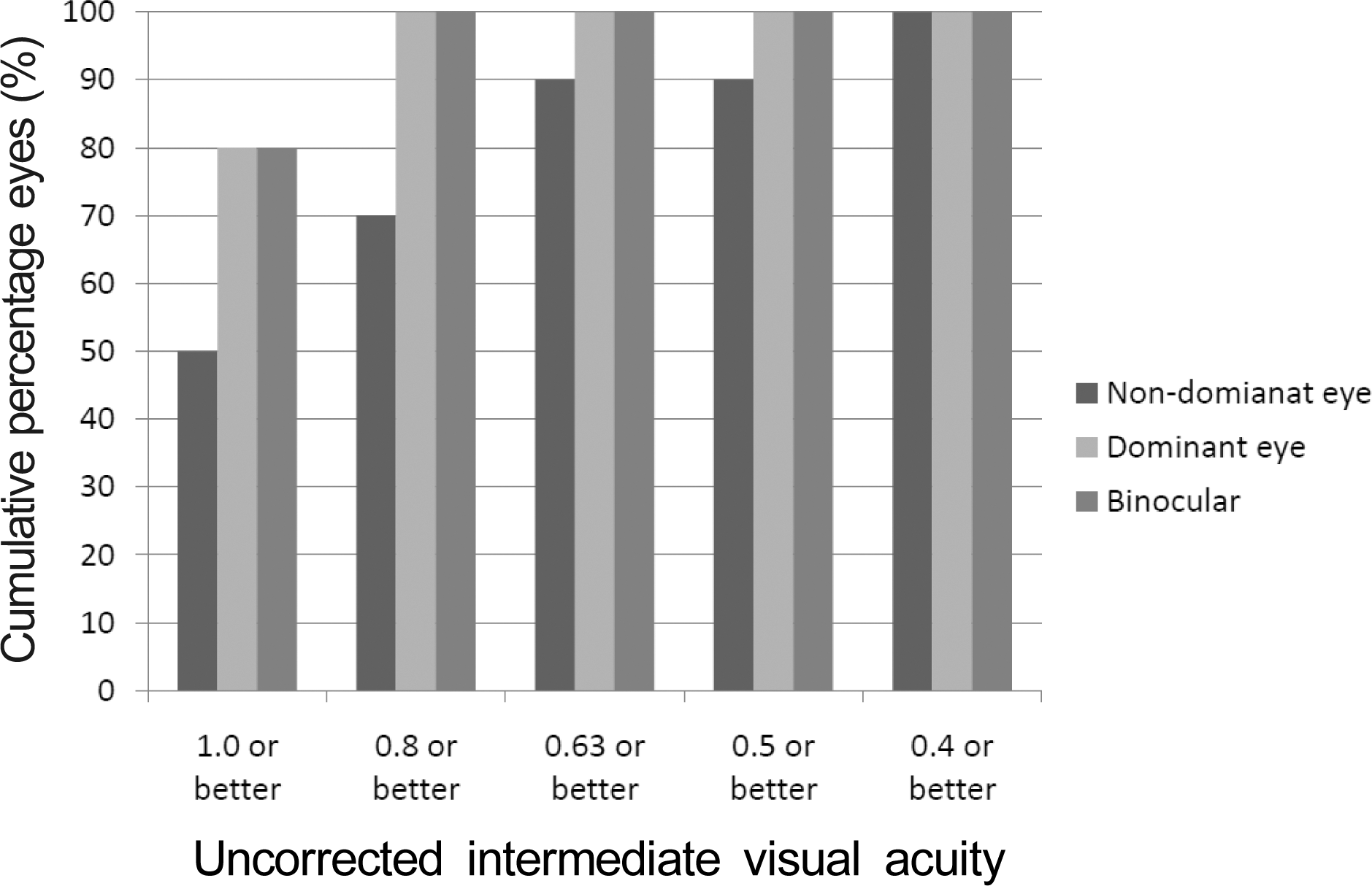
Figure 4.
Histogram showing the accuracy to the intended spherical equivalent refraction 12 months after treatment.
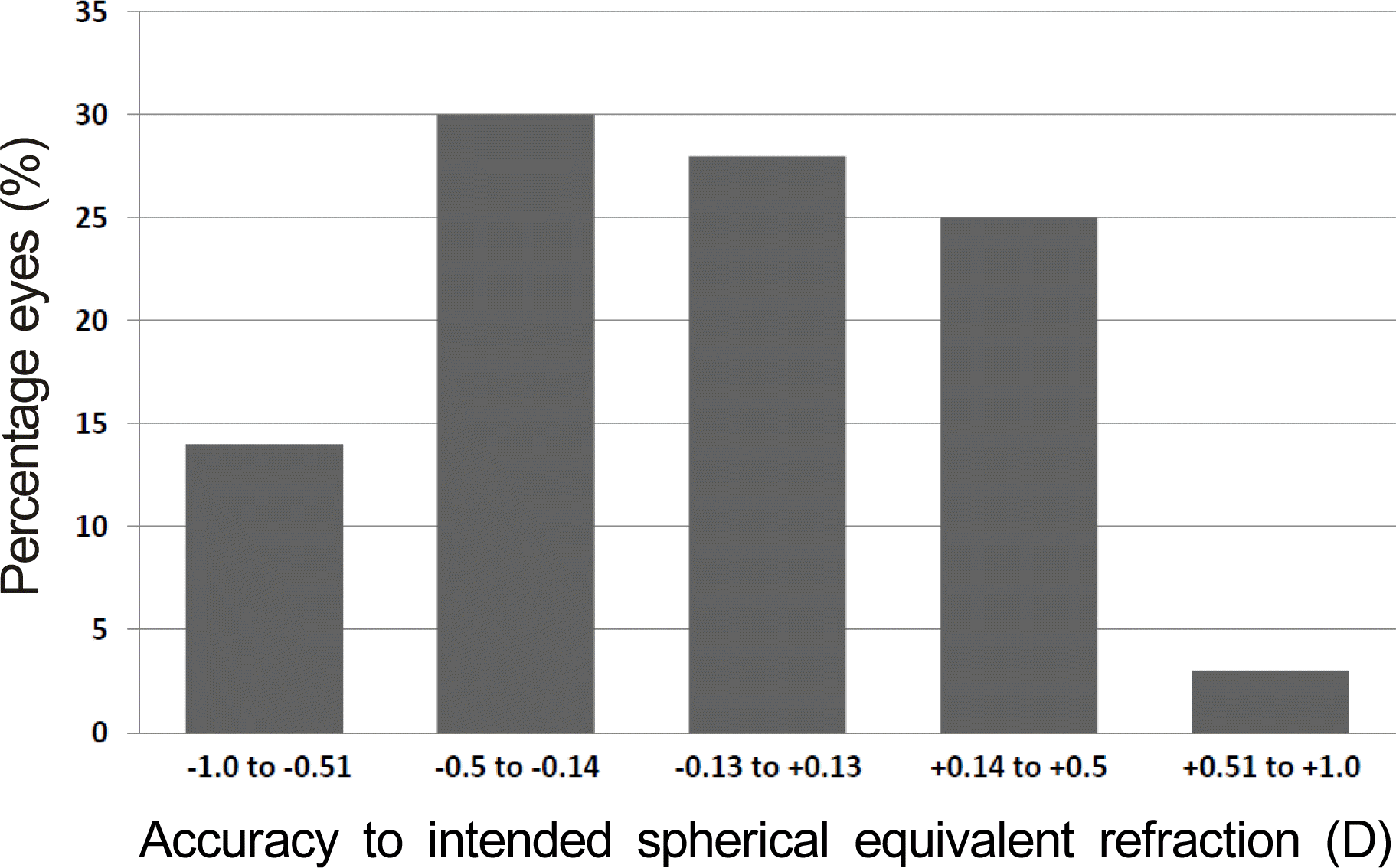
Figure 5.
Cumulative histogram for the distribution of the de-focus equivalent 12 months after treatment.
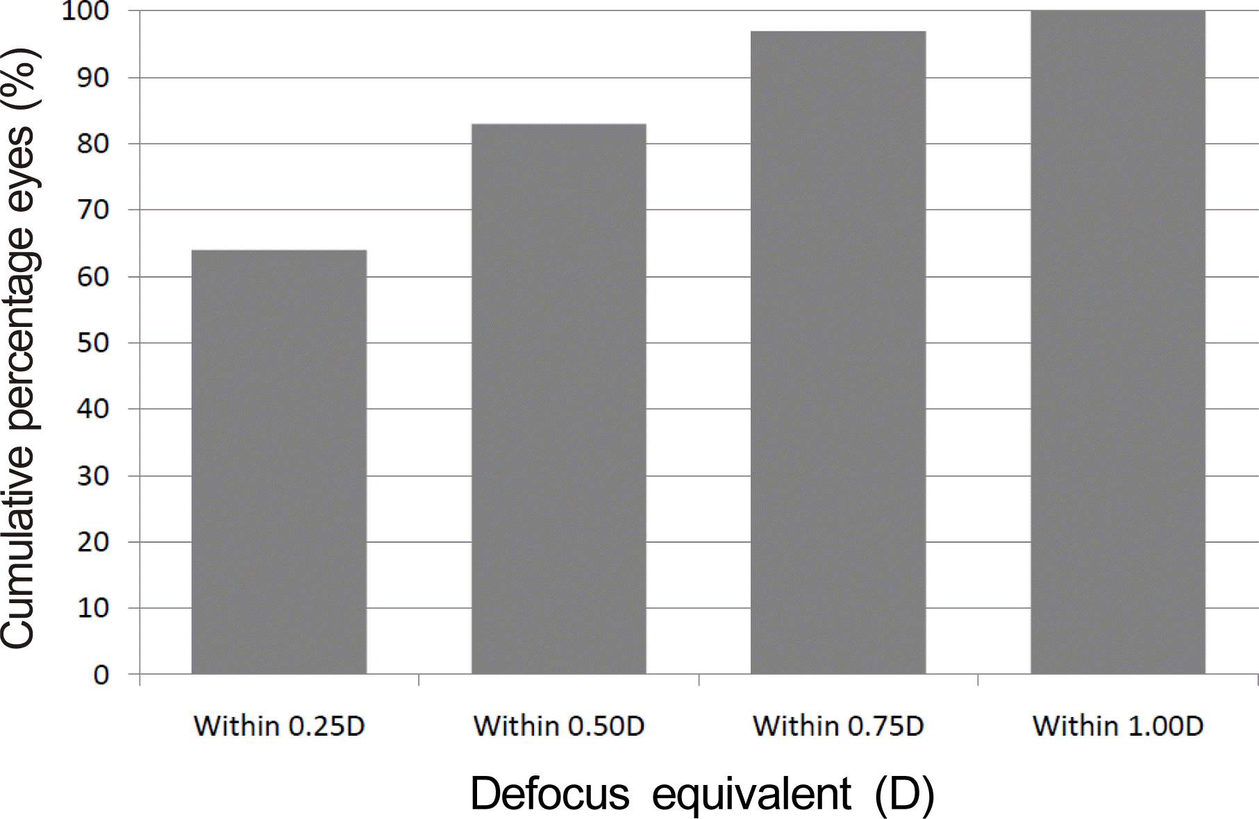
Figure 6.
Perioperative changes in mean spherical equivalent refraction over 12 months after treatment.
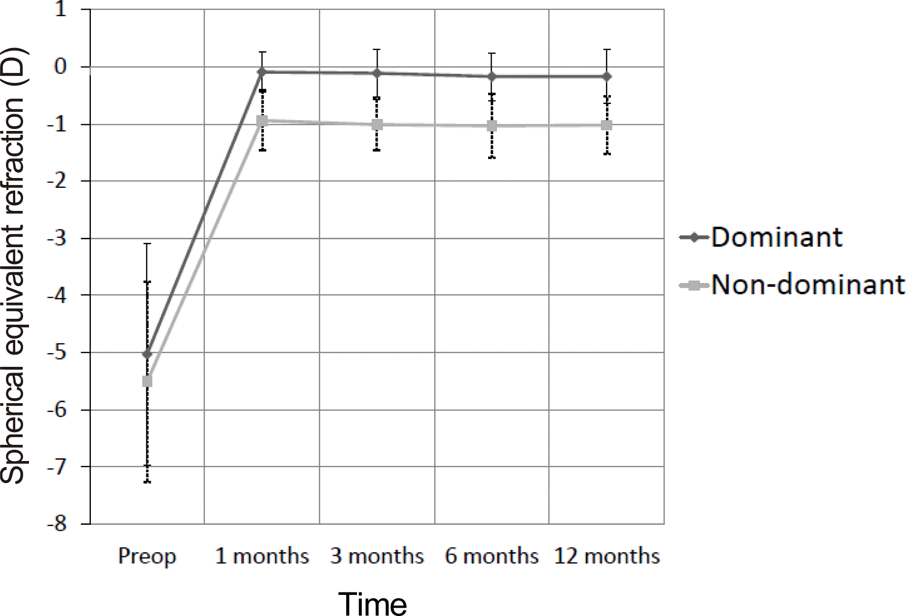
Table 1.
Preoperative patient characteristics


 PDF
PDF ePub
ePub Citation
 Print
Print


 XML Download