Abstract
Purpose
To analyze the features of corneal tissue in patients with posterior polymorphous corneal dystrophy (PPMD) using in vivo confocal microscopy (IVCM).
Case summary
Three patients with clinically diagnosed PPMD were examined using IVCM. Cross-sectioned corneal images of the corneal epithelium, Bowman's layer, stromal layer, Descemet's membrane, and endothelium were evaluated. IVCM demonstrated a depressed crater-like lesion, hyper-dense streak-like lesion, and surface irregularity of the corneal endothelium. Endothelial hypo-reflective vesicular and band-like lesions were also found. Pleomorphism and polymegathism were present with guttae and hyper-reflective endothelial nuclei.
Go to : 
References
1. Vanathi M Tand, on R, Sharma N, et al. In vivo slit scanning con- focal microscopy of normal corneas in Indian eyes. Indian J Ophthalmol. 2003; 51:225–30.
2. Cibis GW, Krachmer JA, Phelps CD, Weingeist TA. The clinical spectrum of posterior polymorphous dystrophy. Arch Ophthalmol. 1977; 95:1529–37.

3. Krachmer JH. Posterior polymorphous corneal dystrophy: a dis- ease characterized by epithelial-like endothelial cells which influ- ence management and prognosis. Trans Am Ophthalmol Soc. 1985; 83:413–75.
4. Park IC, Chung SK, Myong YW, Rhee SW. A case of posterior pol- ymorphous dystrophy. J Korean Ophthalmol Soc. 1991; 32:1020–3.
5. Héon E, Greenberg A, Kopp KK, et al. VSX1 : a gene for posterior polymorphous dystrophy and keratoconus. Hum Mol Genet. 2002; 11:1029–36.
6. Biswas S, Munier FL, Yardley J, et al. Missense mutations in COL8A2, the gene encoding the alpha2 chain of type VIII colla- gen, cause two forms of corneal endothelial dystrophy. Hum Mol Genet. 2001; 10:2415–23.
7. Waring GO 3rd, Rodrigues MM, Laibson PR. Corneal dystrophies. II. Endothelial dystrophies. Surv Ophthalmol. 1978; 23:147–68.

8. Laganowski HC, Sherrard ES, Muir MG. The posterior corneal surface in posterior polymorphous dystrophy: a specular micro- scopical study. Cornea. 1991; 10:224–32.
9. Sekundo W, Lee WR, Kirkness CM, et al. An ultrastructural inves- tigation of an early manifestation of the posterior polymorphous dystrophy of the cornea. Ophthalmology. 1994; 101:1422–31.
10. Feil SH, Barraquer J, Howell DN, Green WR. Extrusion of abnor- mal endothelium into the posterior corneal stroma in a patient with posterior polymorphous dystrophy. Cornea. 1997; 16:439–46.
11. Henriquez AS, Kenyon KR, Dohlman CH, et al. Morphologic characteristics of posterior polymorphous dystrophy. A study of nine corneas and review of the literature. Surv Ophthalmol. 1984; 29:139–47.

12. Patel DV, Grupcheva CN, McGhee CN. In vivo confocal micro- scopy of posterior polymorphous dystrophy. Cornea. 2005; 24:550–4.
Go to : 
 | Figure 1.Slit-lamp photographs of the posterior polymorphous corneal dystrophy patient (case 1). Rail-road like bands on the endothelial surface (black arrows). Below the band like lesion, multiple small vesicles (black arrow- heads) are observed. |
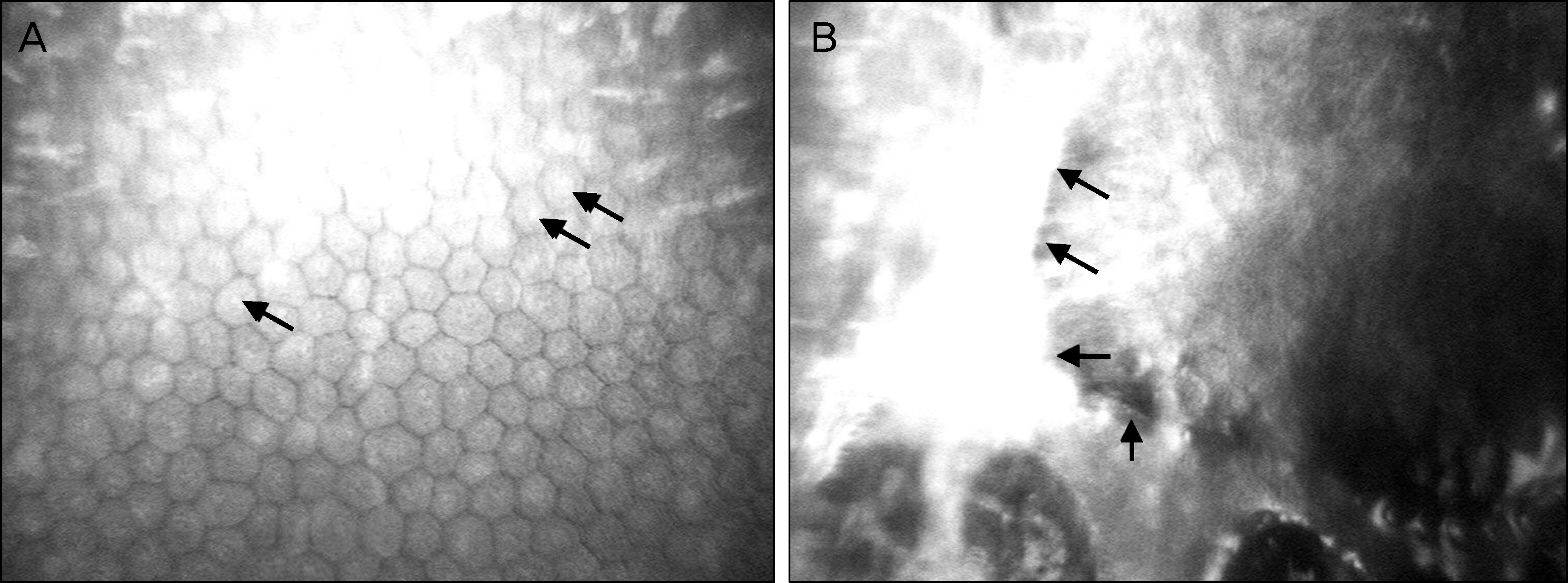
 | Figure 2.In vivo confocal microscopic findings of the posterior polymorphous corneal dystrophy patient (case 1). (A) Hyper-reflective endothelial nuclei (black arrows). (B) Undulating endothelial surface, depressed crater-like lesion and vesicles are noted at the endothelial level (black arrows). |
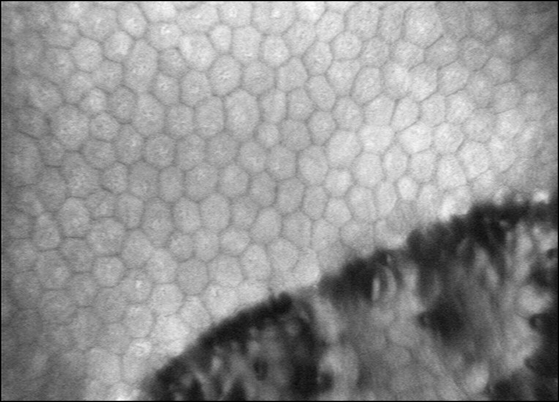
 | Figure 3.In vivo confocal microscopic findings of the posterior polymorphous corneal dystrophy patient (case 2). (A) Endothelial vesicular lesions in curvilinear pattern with associated endothelial pleomorphism and polymegatism. (B) Focal vesicular endothelial lesions (black arrows). (C) Undulating endothelial surface with depressed crater-like lesion and hyper dense streak-like lesion (black arrows). Vesicles and guttata (black arrowheads) are also noted. |
 | Figure 4.In vivo confocal microscopic findings of the posterior polymorphous corneal dystrophy patient (case 3). Endothelial vesicular lesions in curvilinear pattern with associated endothelial pleomorphism and polymegatism. Undulating endothelial surface with depressed crater like lesion is also noted. |


 PDF
PDF ePub
ePub Citation
 Print
Print


 XML Download