

 PDF
PDF ePub
ePub Citation
 Print
Print


Abstract
Purpose
Vasoproliferative tumor of the retina (VPTR) is a histologically benign lesion that can lead to visual loss due to associated complications. Herein, the authors report the clinical presentation, treatment, and prognosis of 3 VPTR cases.
Case summary
Three eyes of 3 patients with VPTR were enrolled in the present study. The patients' fundoscopic feature showed characteristic elevated lesions of the peripheral retina without previous history of ocular disease. The patients included 2 males and 1 female, with an average age of 44.7 years. Fluorescein angiography (FAG) and indocyanine green angiography (ICGA) were helpful in establishing the diagnosis of VPTR. Additionally, 1 patient underwent ultrasonography and 2 patients underwent magnetic resonance imaging (MRI). During the follow-up period, subtenon triamcinolone acetonide injection was performed for 1 patient with macular edema, and cryotherapy was performed for 1 patient with increased peripheral exudation.
Go to : 
References
1. Shields JA, Decker WL, Sanborn GE, et al. Presumed acquired abdominal hemangiomas. Ophthalmology. 1983; 90:1292–300.
2. Jain K, Berger AR, Yucil YH, McGowan HD. Vasoproliferative abdominal of the retina. Eye (Lond). 2003; 17:364–8.
3. Shields CL, Shields JA, Barrett J, De Potter P. Vasoproliferative abdominals of the ocular fundus. Classification and clinical abdominal in 103 patients. Arch Ophthalmol. 1995; 113:615–23.
4. Smeets MH, Mooy CM, Baarsma GS, et al. Histopathology of a vasoproliferative tumor of the ocular fundus. Retina. 1998; 18:470–2.

5. Heimann H, Bornfeld N, Vij O, et al. Vasoproliferative tumours of the retina. Br J Ophthalmol. 2000; 84:1162–9.
6. Irvine F, O'Donnell N, Kemp E, Lee WR. Retinal vasoproliferative tumors: surgical management and histological findings. Arch Ophthalmol. 2000; 118:563–9.
7. Anastassiou G, Bornfeld N, Schueler AO, et al. Ruthenium-106 plaque brachytherapy for symptomatic vasoproliferative tumours of the retina. Br J Ophthalmol. 2006; 90:447–50.
8. Cohen VM, Shields CL, Demirci H, Shields JA. Iodine I 125 abdominal radiotherapy for vasoproliferative tumors of the retina in 30 eyes. Arch Ophthalmol. 2008; 126:1245–51.
9. Saldanha MJ, Edrich C. Treatment of vasoproliferative tumors with photodynamic therapy. Ophthalmic Surg Lasers Imaging. 2008; 39:143–5.
10. Chan RP, Lai TY. Photodynamic therapy with verteporfin for abdominal tumour of the retina. Acta Ophthalmol. 2010; 88:711–2.
11. Yeh S, Wilson DJ. Pars plana vitrectomy and endoresection of a retinal vasoproliferative tumor. Arch Ophthalmol. 2010; 128:1196–9.
12. Kenawy N, Groenwald C, Damato B. Treatment of a abdominal tumour with intravitreal bevacizumab (Avastin). Eye (Lond). 2007; 21:893–4.
13. McAllister IL, Vijayasekaran S, Chen SD, Yu DY. Effect of abdominal acetonide on vascular endothelial growth factor and occludin levels in branch retinal vein occlusion. Am J Ophthalmol. 2009; 147:838–46. 846.e1–2.
14. Hayashi K, Hayashi H. Intravitreal versus retrobulbar injections of triamcinolone for macular edema associated with branch retinal vein occlusion. Am J Ophthalmol. 2005; 139:972–82.
15. Byun YS, Park YH. Complications and safety profile of posterior subtenon injection of triamcinolone acetonide. J Ocul Pharmacol Ther. 2009; 25:159–62.
Go to :
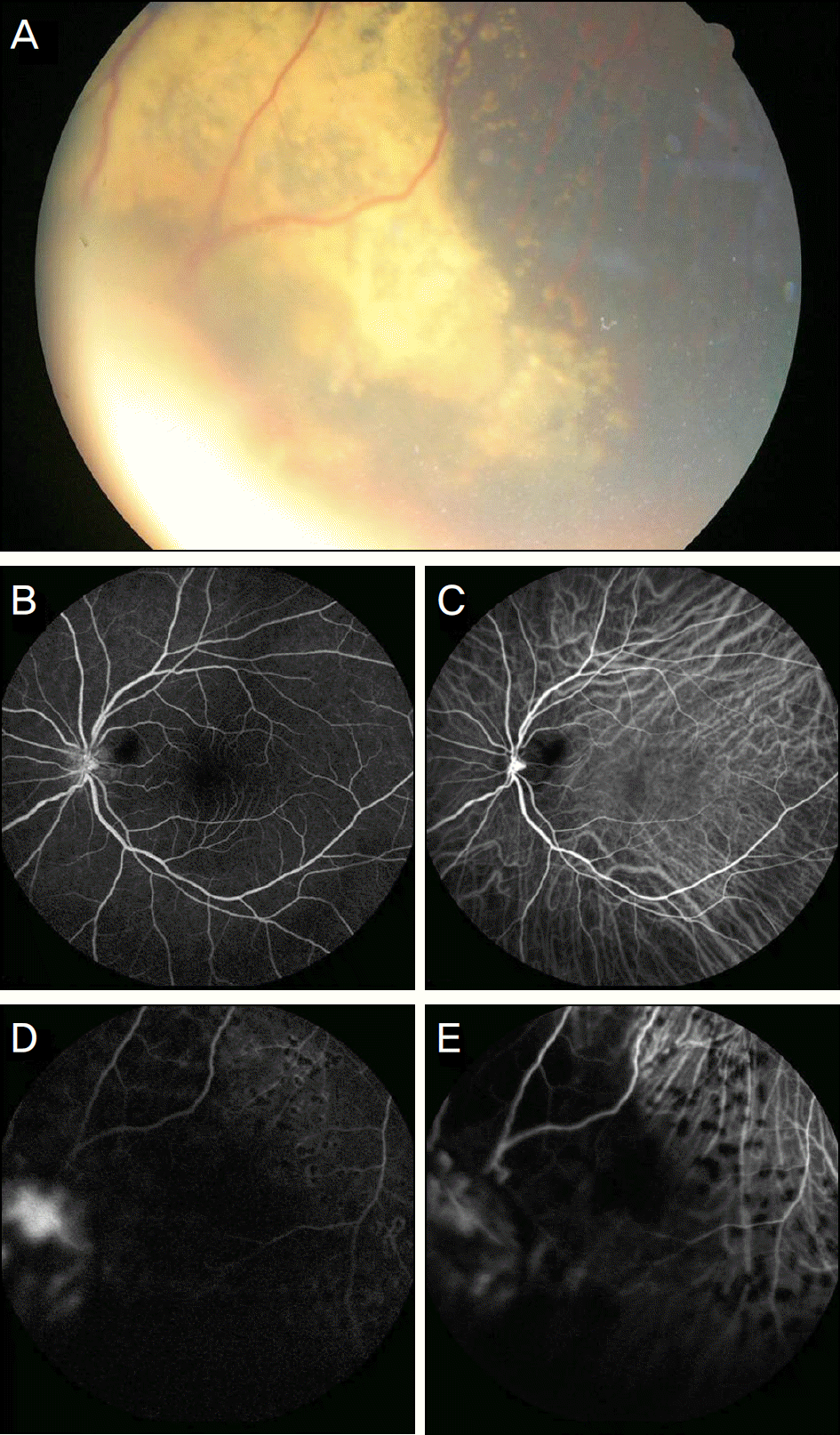
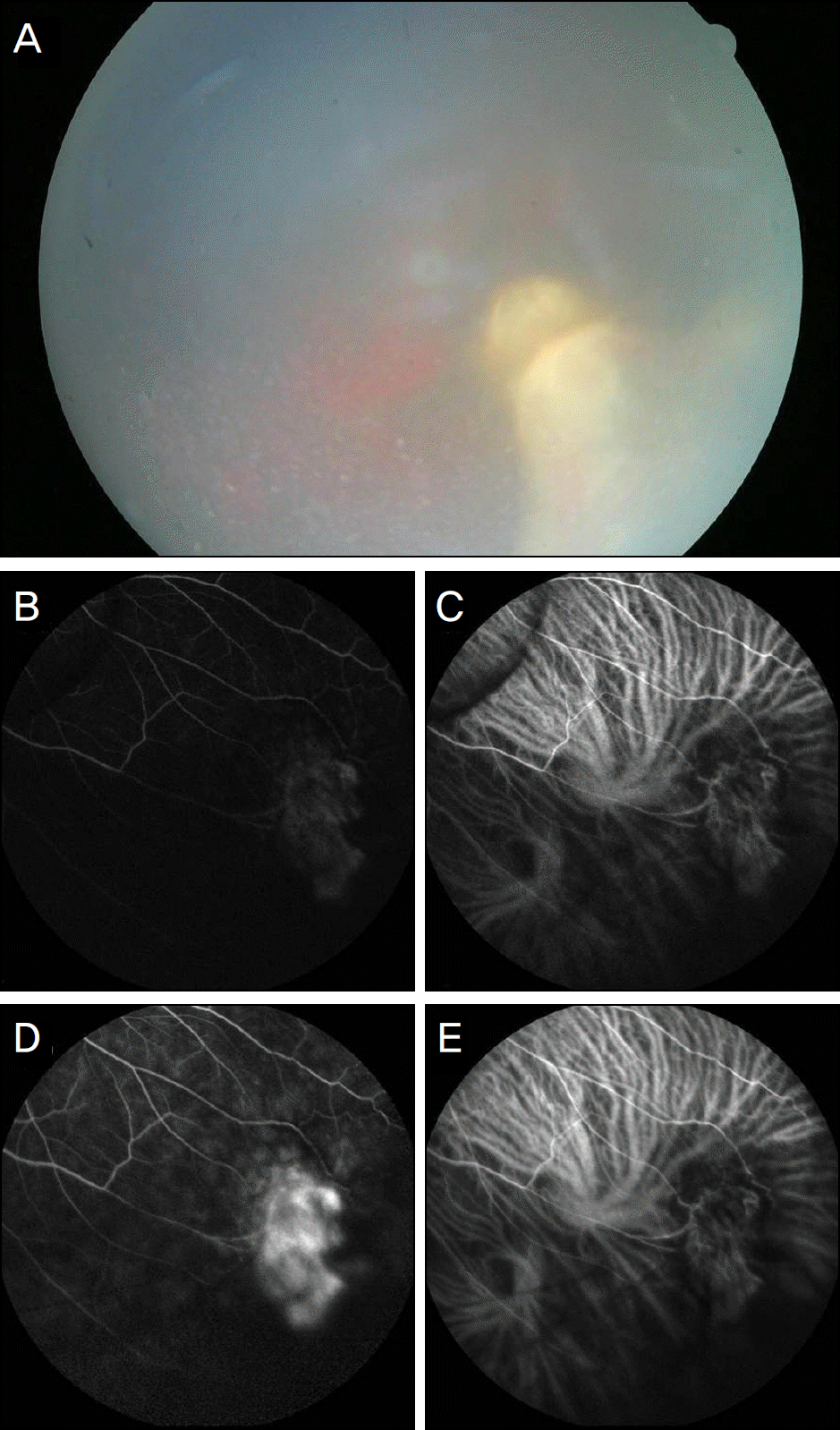 | Figure 1.(A) Homogenous creamy yellow colored fine mass in inferotemporal peripheral retina. (B) and (D) FAG finding. Dilated and obscured vessels with hyperfluorescence on mass lesion. There were neither abnormal feeding vessels nor dilated vessels in surrounding mass lesion. (C) and (E) ICGA finding. Blocked fluorescence associated with tumor showing no abnormal choroidal vessels. |
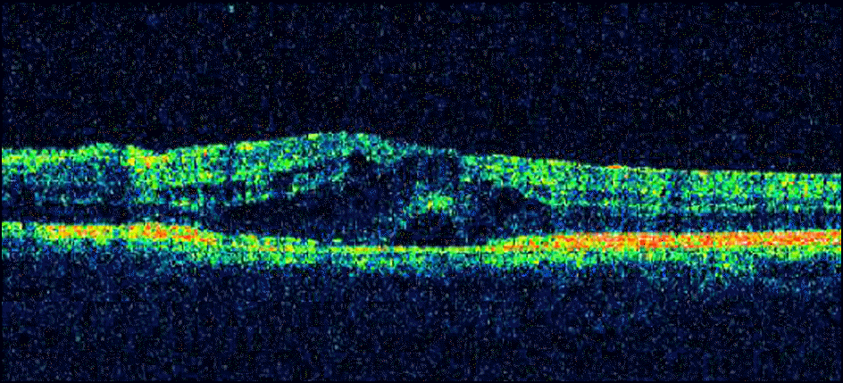 | Figure 2.Optical coherence tomographic finding. Maular edema with thin epiretinal membrane. |
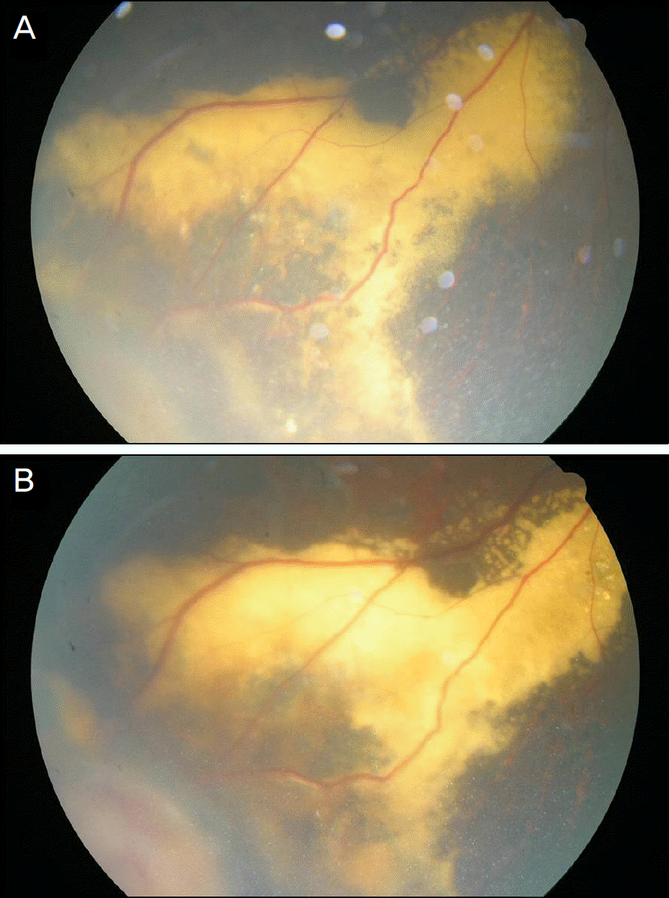
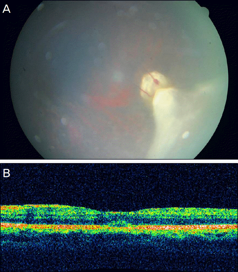 | Figure 3.(A) Well visualized retinal mass through relative clear vitreous cavity 1 month after subtenon's triamcinolone aceteonide injection. (B) Optical coherence tomographic finding. Absorbed macular edema one month after subtenon's triamcinolone aceteonide injection. |
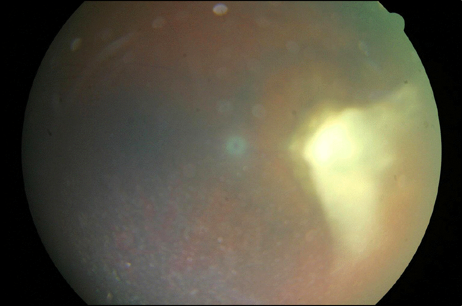
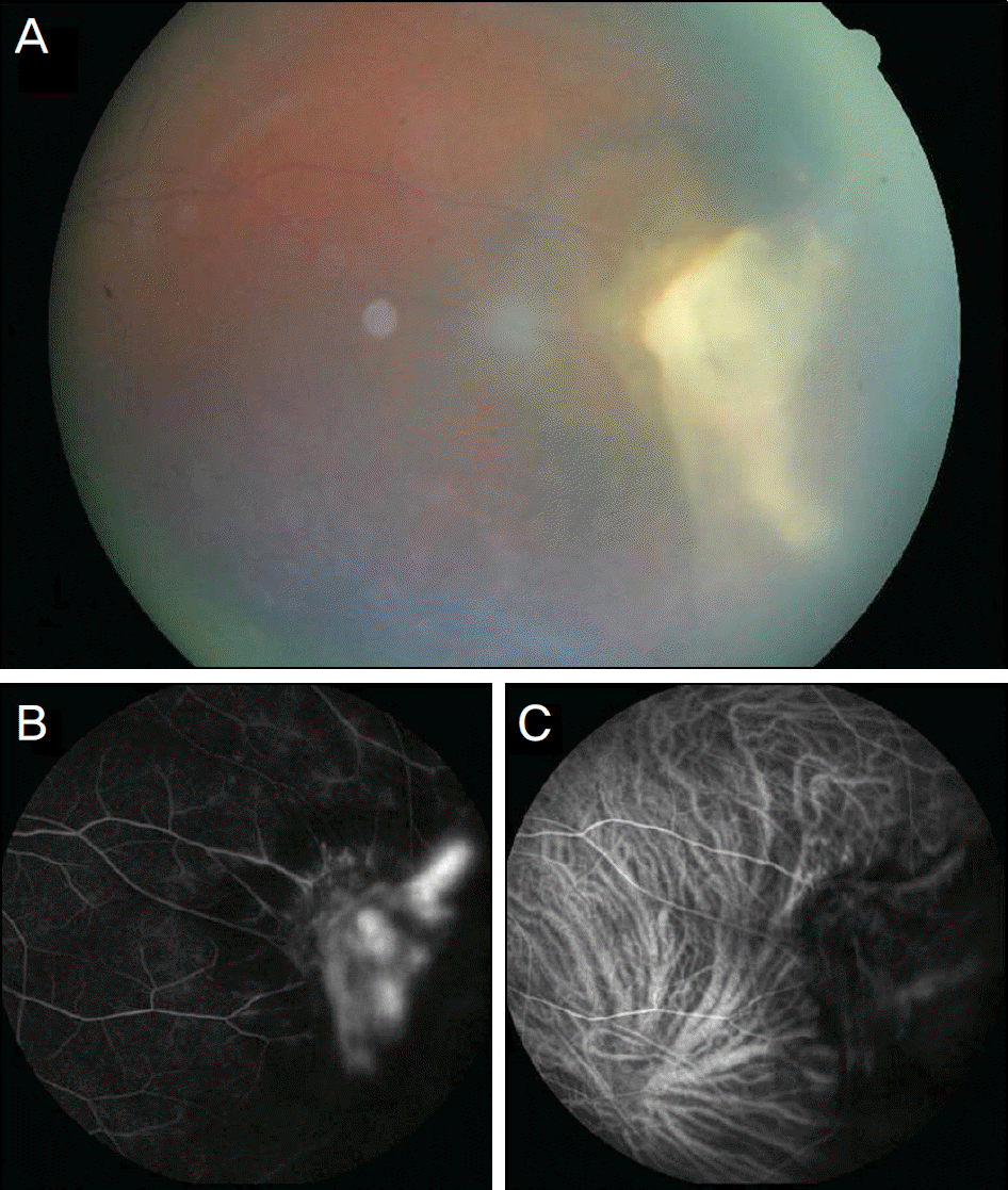 | Figure 4.(A) Homogenous creamy yellow colored mass in inferotemporal peripheral retina. (B) FAG finding. Dilated and obscure vessels with hyperfluorescence on mass lesion. There were neither abnormal feeding vessels nor dilated vessels in surrounding mass lesion. (C) ICGA finding. Blocked fluorescence associated with tumor showing no abnormal choroidal vessels. |
 | Figure 6.(A) Homogenous creamy yellow colored fine mass with surrounding exudates and photocoagulation scar in inferonasal peripheral retina. (B) and (C) Normal macular appearance. (D) FAG finding. Dilated and obscured vessels with hyperfluorescence on mass lesion and visible photo-coagulation scar. There were neither abnormal feeding vessels nor dilated vessels in surrounding mass lesion. (E) ICGA finding. Blocked fluorescence associated with subretinal exudates and photocoagulation scar showing no abnormal choroidal vessels. |
 XML Download