Abstract
Purpose
Retinal detachment (RD) complicated in acute retinal necrosis (ARN) is difficult to be treated and a main cause of blindness. The factors associated with RD in ARN were investigated.
Methods
Patients with ARN who were diagnosed and treated from Jan, 2008 to Dec, 2012 were reviewed retrospectively. The eyes were classified into the group I without RD, and the group II with RD. Early vitrectomy, history of ARN in the other eye, extent of necrosis, symptom duration and intravitreal injection of anti-viral drug were evaluated.
Results
Of 22 eyes of 20 patients, 11 eyes were included in each group. Symptom duration of 8.0 days in the group I was shorter than 15.8 days in the group II (p = 0.005). There were no macular involvement at initial exam in the group I and 5 eyes (45%) in the group II (p = 0.017). Five eyes (45%) in the group I and 0 eye (0%) in the group II had history of ARN in the other eye (p = 0.017). Six eyes (55%) in the group I and 1 eye (9%) in the group II underwent early vitrectomy (p = 0.031). Age, baseline visual acuity, and intravitreal injection of antiviral agent were not related to RD (p = 0.294-0.699).
Go to : 
References
1. Fisher JP, Lewis ML, Blumenkranz M, et al. The acute retinal ne- crosis syndrome. Part 1: clinical manifestations. Ophthalmology. 1982; 89:1309–16.
2. Clarkson JG, Blumenkranz MS, Culbertson WW, et al. Retinal de- tachment following the acute retinal necrosis syndrome. Ophthalmology. 1984; 91:1665–8.
3. Culbertson WW, Blumenkranz MS, Haines H, et al. The acute reti- nal necrosis syndrome: Histopathology and etiology. Ophthalmology. 1982; 89:1317–25.
4. Iwahashi-Shima C, Azumi A, Ohguro N, et al. Acute retinal ne- crosis: factors associated with anatomic and visual outcomes. Jpn J Ophthalmol. 2013; 57:98–103.
5. Hillenkamp J, Nölle B, Bruns C, et al. Acute retinal necrosis: clin- ical features, early vitrectomy, and outcomes. Ophthalmology. 2009; 116:1971–5.e2.
6. Luo YH, Duan XC, Chen BH, et al. Efficacy and necessity of pro- phylactic vitrectomy for acute retinal necrosis syndrome. Int J Ophthalmol. 2012; 5:482–7.
7. Wong R, Pavesio CE, Laidlaw DA, et al. Acute retinal necrosis: The effects of intravitreal foscarnet and virus type on outcome. Ophthalmology. 2010; 117:556–60.
8. Holland GN. Standard diagnostic criteria for the acute retinal ne- crosis syndrome. Executive Committee of the American Uveitis Society. Am J Ophthalmol. 1994; 117:663–7.
9. Holland GN, Buhles WC Jr, Mastre B, Kaplan HJ. A controlled retrospective study of ganciclovir treatment for cytomegalovirus retinopathy. Use of a standardized system for the assessment of dis- ease outcome. Arch Ophthalmol. 1989; 107:1759–66.
10. Ishida T, Sugamoto Y, Sugita S, Mochizuki M. Prophylactic vi- trectomy for acute retinal necrosis. Jpn J Ophthalmol. 2009; 53:486–9.
11. Jung JY, Lee TG. Vitrectomy and silicone oil tamponade to prevent retinal detachment in severe acute retinal necrosis syndrome. J Korean Ophthalmol Soc. 2008; 49:519–24.

12. Yang JW, Kim WJ, Park YH. Two cases of acute retinal necrosis treated with systemic antiviral drugs and intravitreal antiviral injections. J Korean Ophthalmol Soc. 2009; 50:794–9.

13. Coen DM, Fleming HE Jr, Leslie LK, Retondo MJ. Sensitivity of arabinosyladenine-resistant mutants of herpes simplex virus to other antiviral drugs and mappingof drug hypersensitivity mutations to the DNA polymerase locus. J Virol. 1985; 53:477–88.
Go to : 
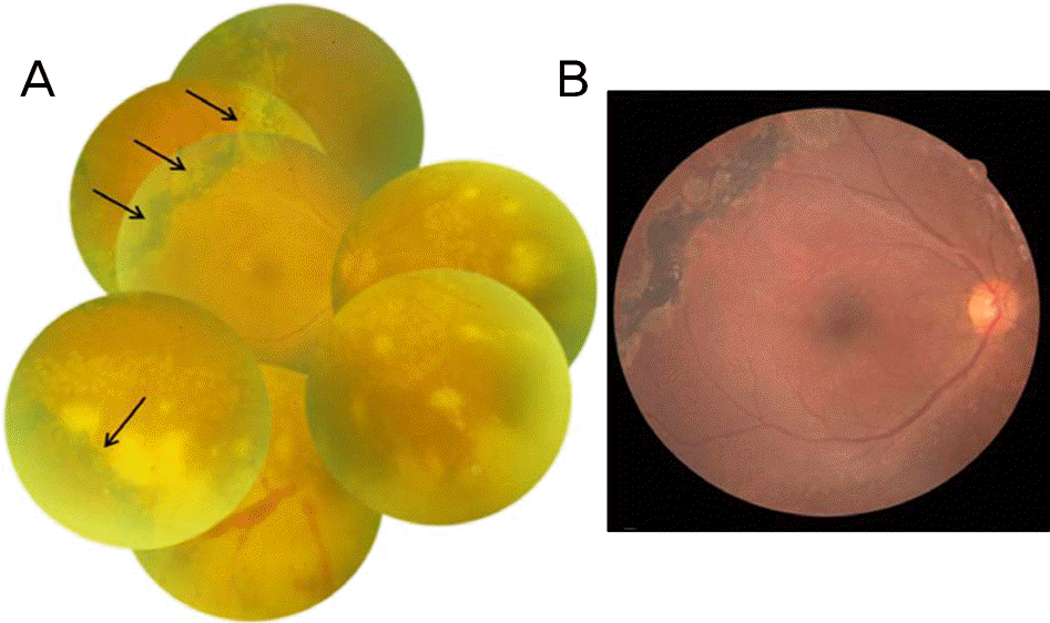
 | Figure 1.Group I, patient 5. (A) Two days after systemic antiviral and laser treatment, fundus photo shows that retinal necrosis did not extend to the macula. Pigmented laser scar (black arrow) by previous treatment several years ago is noted. (B) Five months after early vitrectomy, there is no retinal detachment, vitreous inflammation, or retinal necrosis. |
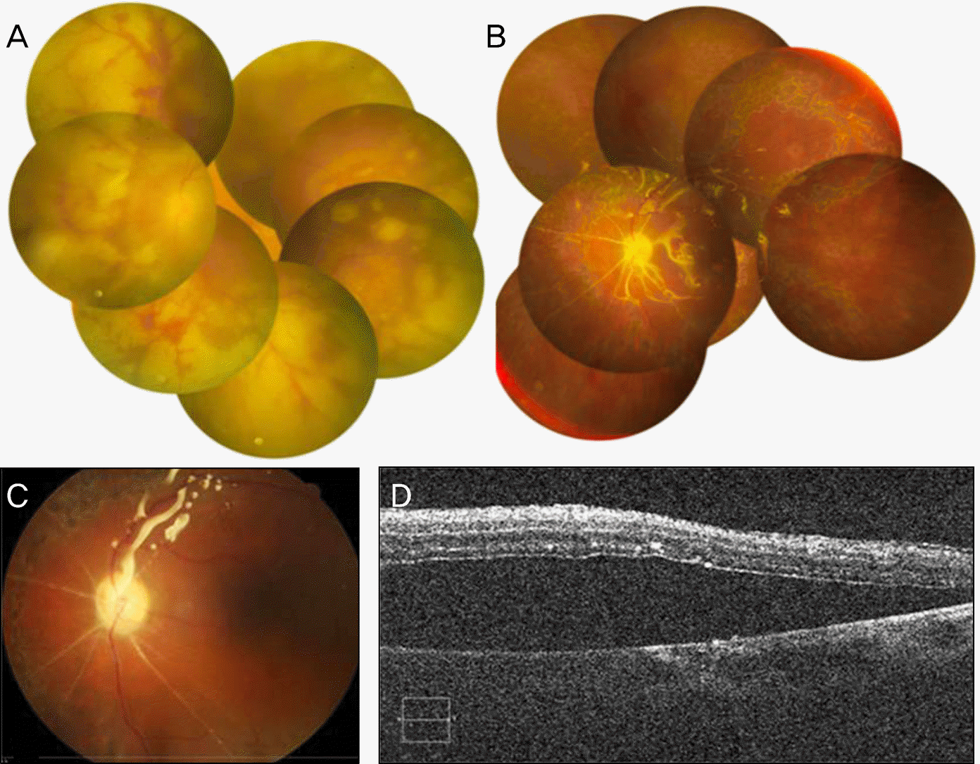
 | Figure 2.Group II, patient 12. (A) Two days after systemic antiviral and laser treatment, fundus photo shows that the macula was involved by retinal necrosis. (B) Ten days after early vitrectomy, vitreous cavity was filled with silicone oil and the retina is attached without necrotic lesion. (C) Fifty days after early vitrectomy, although it is filled with the tamponade, subretinal fluid is note. (D) Optical coherence tomography demonstrates presence of subretinal fluid. |
Table 1.
Demographic characteristics of the patients
Table 2.
Patient data


 PDF
PDF ePub
ePub Citation
 Print
Print


 XML Download