Abstract
Purpose
In this study we evaluated the changes in tear film after primary pterygium operation in patients with pterygium.
Methods
We investigated 43 eyes of 42 subjects who showed successful results 3 months after pterygium operation performed by one surgeon. The changes in tear film thickness, tear break-up time (BUT), Schirmer I test, and ocular surface disease index (OSDI) were evaluated. All values were compared before and after surgery.
Results
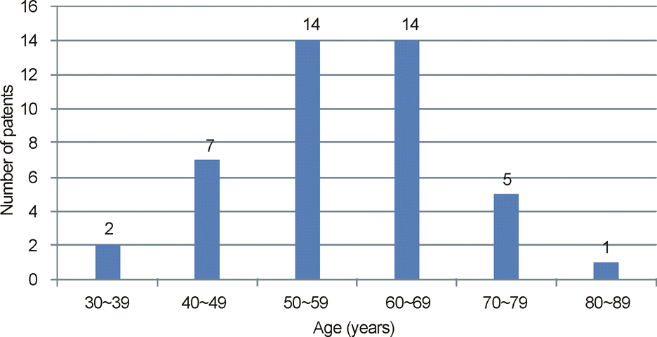
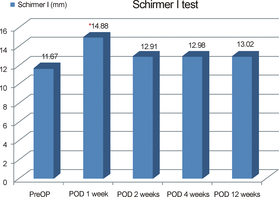
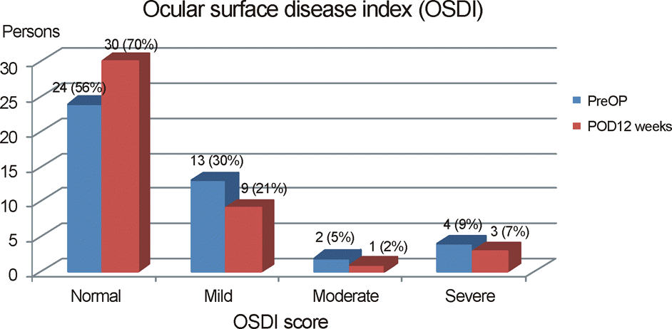
The mean age was 58.0 ± 11.1 years (34-81 years). Preoperative tear film thickness, tear BUT, and Schirmer I test in eyes which underwent pterygium operation were 21.53 ± 5.93 μm, 4.84 ± 2.21 seconds, and 11.67 ± 6.75 mm, respectively. Three months after the operation, the respective values were 24.23 ± 4.19 μm (p < 0.05), 5.81 ± 1.89 seconds (p < 0.05), and 13.02 ± 7.54 mm (p = 0.094). Tear film thickness and BUT score increased significantly after pterygium operation. There was no statistically significant difference in Schirmer I test, before and 3 months after pterygium operation. The subjective parameter (OSDI) improved 3 months after pterygium operation (p = 0.015).
Go to : 
References
1. Mutlu FM, Sobaci G, Tatar T, Yildirim E. A comparative study of recurrent pterygium surgery: limbal conjunctival autograft trans- plantation versus mitomycin c with conjunctival flap. Ophthalm- ology. 1999; 106:817–21.
2. Ang LP, Chua JL, Tan DT. Current concepts and techniques in pterygium treatment. Curr Opin Ophthalmol. 2007; 18:308–13.

3. Corneo MT, Di Girolamo N, Wakefield D. The pathogenesis of pterygia. Curr Opin Ophthalmol. 1999; 10:282–8.
4. Di Girolamo N, Chui J, Corneo MT, Wakefield D. Pathogenesis of pterygia: role of cytokines, growth factor, and matrix metallo- proteinase. Prog Retin Eye Res. 2004; 23:195–228.
5. Coroneo MT. Pterygium as an early indicator of ultraviolet in- solation: a hypothesis. Br J Opthalmol. 1993; 77:734–9.
6. Ergin A, Bozdogan O. Study on tear function abnormality in pterygium. Ophthalmologica. 2001; 215:204–8.

7. Li M, Zhang M, Lin Y, et al. Tear function and goblet cell density after pterygium excision. Eye (Lond). 2007; 24:224–8.

8. Ischioka M, Shimmura S, Yagi Y, Tsubota K. Pterygium and dry eye. Ophthalmologica. 2001; 215:209–11.
9. Wang S, Jiang B, Gu Y. Changes of tear film function after ptery- gium operation. Ophthalmic Res. 2011; 45:210–5.
10. Oh HJ, Park YG, Yoon KC. Changes of ocular surface and tear film in patients with pinguecula and pterygium. J Korea Ophthalmol Soc. 2006; 47:717–24.
11. Zhuang H, Zhou X, Xu J. A novel method for pachymetry mapping of human precorneal tear film using pentacam with fluorescein. Invest Ophthalmol Vis Sci. 2010; 51:156–9.

12. Schiffman RM, Christianson MD, Jacobsen G, et al. Reliability and validity of the Ocular Surface Disease Index. Arch Ophthalmol. 2000; 118:615–21.

13. Miller KL, Walt JG, Mink DR, et al. Minimal clinically important difference for the ocular surface disease index. Arch Ophthalmol. 2010; 128:94–101.

14. Goldberg L, David R. Pterygium and its relationship to the dry eye in the Bantu. Br J Ophthalmol. 1976; 60:720–1.

15. Biedner B, Biger Y, Rothkoff L, Sachs U. Pterygium and basic tear secretion. Ann Ophthalmol. 1979; 11:1235–6.
16. Kadayifçilar SC, Orhan M, Irkeç M. Tear function in patients with pterygium. Acta Ophthalmol Scand. 1998; 76:176–9.
17. Holly FJ. Physical chemistry of the normal and disordered tear film. Trans Ophthalmol Soc U K. 1985; 104(Pt 4):374–80.
18. Marzeta M, Toczolowski J. [Study of mucin of tear film in patients with pterygium]. Klin Oczna. 2003; 105:60–2.
19. Nakagami T, Watanabe I, Murakami A, et al. Expression of stem cell factor in pterygium. Jpn J Ophthalmol. 2000; 44:193–7.

20. Tseng SC. Evaluation of the ocular surface in dry-eye conditions. Int Ophthalmol Clin. 1994; 34:57–69.

21. Johnson ME, Murphy PJ. Changes in the tear film and ocular surface from dry eye syndrome. Prog Retin Eye Res. 2004; 23:449–74.

22. Rajiv SM, Mithal S, Sood AK. Pterygium and dry eye–a clinical correlation. Ind J Ophthalmol. 1991; 38:15–6.
23. Marzeta M, Toczolowski J. Study of mucin of tear film in patients with pterygium. Klin Oczna. 2003; 105:60–2.
Go to : 
 | Figure 2.Fluorescent tear film can be visualized by Scheimpflug imaging. Pentacam system generates a color-coded pachymetry map of tear film by comparing two corneal pachymetry maps of each eye. (A) Corneal pachymetry map after flurescein installation. (B) Corneal pachymetry map before flurescein installation. (C) Pachymetry map of tear flim obtained by substracting map B from map A. |
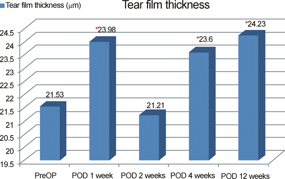 | Figure 3.Changes in tear film thickness before and after pterygium surgery at 1, 2, 4, 12 weeks postoperatively. The postoperative tear film thickness is statistically significant being thicker than the preoperative one except at postoperative 2 weeks. *p < 0.05: Statistically significant. |
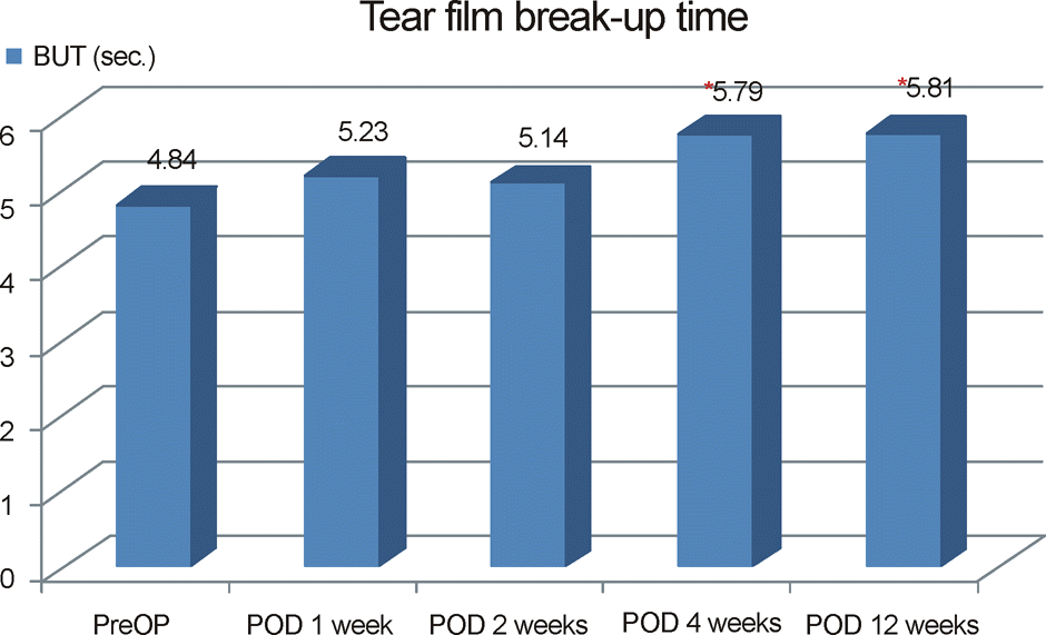 | Figure 4.Changes in tear film break-up time before and after pterygium surgery at 1, 2, 4, 12 weeks postoperatively. The postoperative tear film break-up time is significantly longer than the preoperative after postoperative 4 weeks. *p < 0.05: Statistically significant. |


 PDF
PDF ePub
ePub Citation
 Print
Print


 XML Download