

 PDF
PDF ePub
ePub Citation
 Print
Print


Abstract
Purpose
To report a case of IgG4-related sclerosing disease involving the eyelid in an idiopathic sclerosing myositis patient.
Case summary
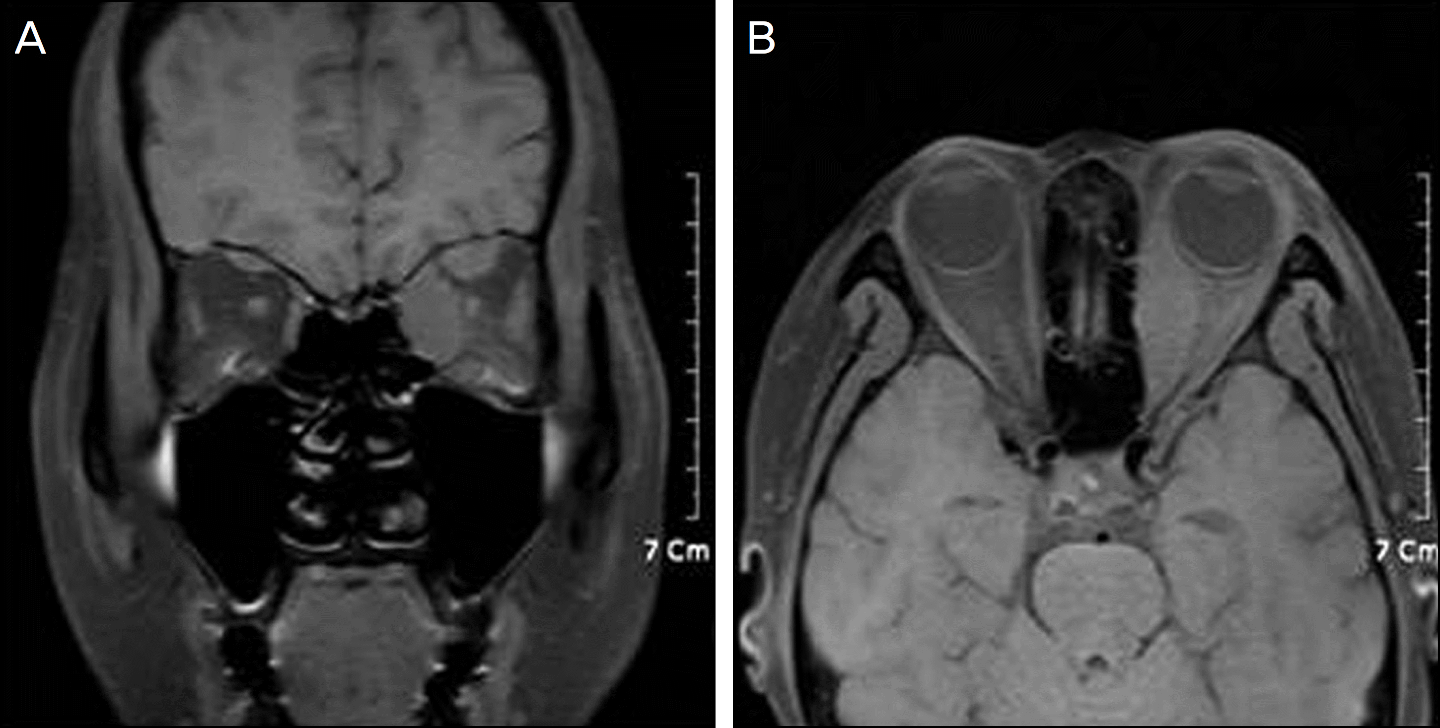
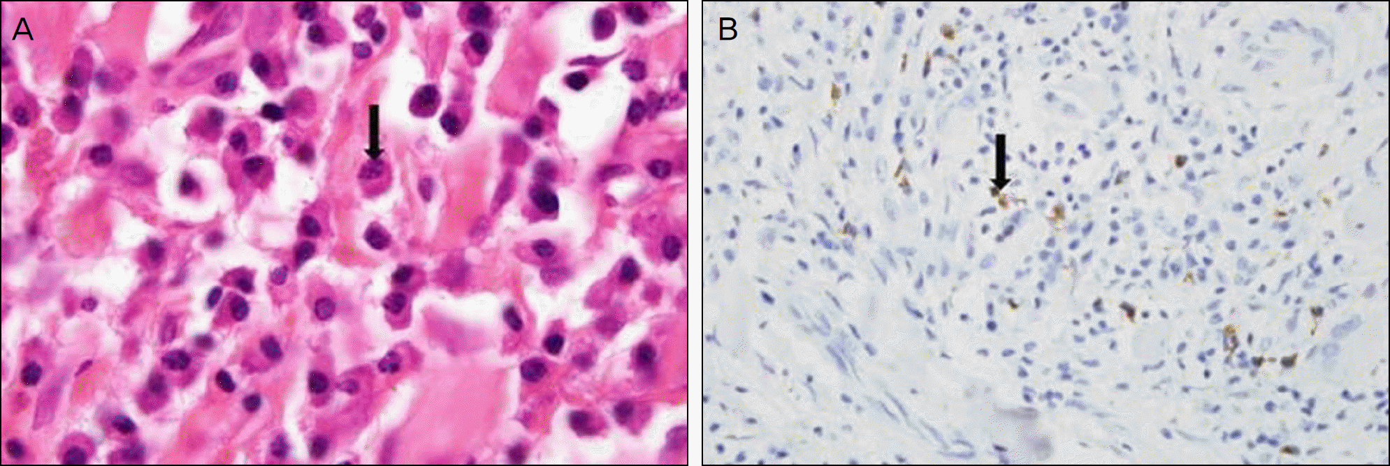
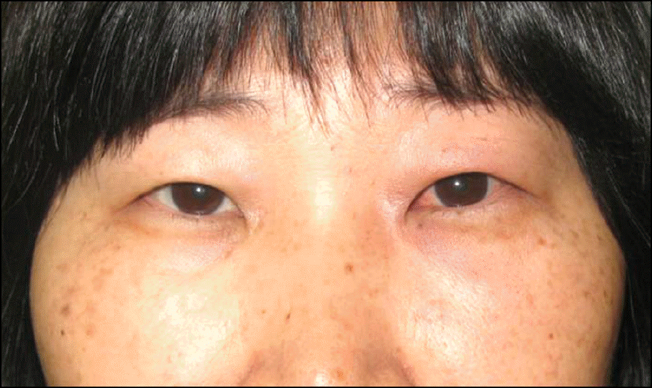
A 51-year-old woman presented with swelling, redness, and tenderness of the left lower eyelid of 1 month duration had taken an immunosuppressant for idiopathic sclerosing myositis. Eye movements showed limitation all directions but there was no exophthalmos. A palpable mass was noted in the left lower eyelid. The left extraocular muscles were hypertrophied but the lacrimal gland was normal on orbital magnetic resonance imaging. IgG4-related sclerosing disease was confirmed by immunostained biopsy from the left lower eyelid, showing infiltration of IgG4-positive lymphoplasmacytic cells. The patient was given oral steroids and an immunosuppressant and the symptoms did not recur for at least 7 months.
Conclusions
IgG4-related sclerosing disease involving ocular adnexa usually consists of bilateral lacrimal gland involvement. Additionally, the orbital soft tissue involvement without dacryoadenitis is rare. The authors of the present study report a case of IgG4-related sclerosing disease involving the left lower eyelid in an idiopathic sclerosing myositis patient and should be considered in the differential diagnosis of eyelid masses.
References
1. Plaza JA, Garrity JA, Dogan A, et al. Orbital inflammation with IgG4-positive plasma cells: manifestation of IgG4 systemic disease. Arch Ophthalmol. 2011; 129:421–8.
2. Kim KE, Lee MJ, Kim NJ, et al. Three cases of Hyper-IgG4 abdominal involving ocular adnexa. J Korean Ophthalmol Soc. 2010; 51:1133–8.
3. Mehta M, Jakobiec F, Fay A. Idiopathic fibroinflammatory disease of the face, abdominals, and periorbital membrane with immunoglobulin G4-positive plasma cells. Arch Pathol Lab Med. 2009; 133:1251–5.
4. Kamisawa T, Funata N, Hayashi Y, et al. A new clinicopathological entity of IgG4-related autoimmune disease. J Gastroenterol. 2003; 38:982–4.

5. Kamisawa T, Okamoto A. IgG4-related sclerosing disease. World J Gastroenterol. 2008; 14:3948–55.
6. Cheuk W, Yuen HK, Chan JK. Chronic sclerosing dacryoadenitis: part of the spectrum of IgG4-related sclerosing disease? Am J Surg Pathol. 2007; 31:643–5.
7. Khosroshahi A, Stone JH. A clinical overview of IgG4-related abdominalic disease. Curr Opin Rheumatol. 2011; 23:57–66.
8. Espinoza GM. Orbital inflammatory pseudotumors: etiology, abdominalial diagnosis, and management. Curr Rheumatol Rep. 2010; 12:443–7.
9. Zen Y, Kitagawa S, Minato H, et al. IgG4-positive plasma cells in inflammatory pseudotumor (plasma cell granuloma) of the lung. Hum Pathol. 2005; 36:710–7.
10. Higashiyama T, Nishida Y, Ugi S, et al. A case of extraocular abdominal swelling due to IgG4-related sclerosing disease. Jpn J Ophthalmol. 2011; 55:315–7.
11. Sato Y, Ohshima K, Ichimura K, et al. Ocular adnexal IgG4-related disease has uniform clinicopathology. Pathol Int. 2008; 58:465–70.
12. Kamisawa T, Tu Y, Nakajima H, et al. Usefulness of biopsying the major duodenal papilla to diagnose autoimmune pancreatitis: a abdominal study using IgG4-immunostaining. World J Gastroenterol. 2006; 12:2031–3.
13. Sato Y, Takata K, Ichimura K, et al. IgG4-producing marginal zone B-cell lymphoma. Int J Hematol. 2008; 88:428–33.
14. Sandanayake NS, Church NI, Chapman MH, et al. Presentation and Mmanagement of Ppost-treatment Rrelapse in Aautoimmune Ppancreatitis/Iimmunoglobulin G4-Aassociated Ccholangitis. Clin Gastroenterol Hepatol. 2009; 7:1089–96.
15. Naitoh I, Nakazawa T, Ohara H, et al. Autoimmune pancreatitis abdominal with various extrapancreatic lesions during a long-term clinical course successfully treated with azathioprine and abdominal maintenance therapy. Intern Med. 2009; 48:2003–7.
16. Matsuo T, Ichimura K, Sato Y, et al. Immunoglobulin G4 (IgG4)-positive or -negative ocular adnexal benign lymphoid lesions in abdominal to systemic involvement. J Clin Exp Hematop. 2010; 50:129–42.
17. Ishii S, Shishido F, Miyajima M, et al. Whole-body gallium-67 scintigraphic findings in IgG4-related disease. Clin Nucl Med. 2011; 36:542–5.
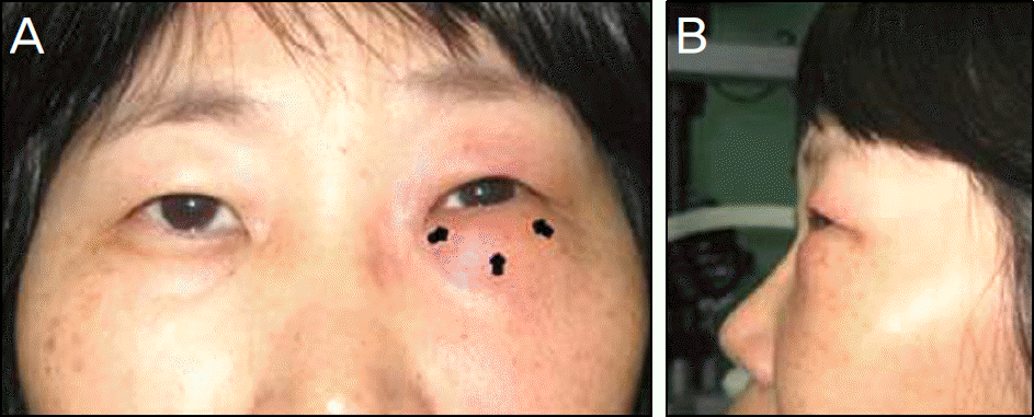
Figure 2.
(A, B) Orbital T1-weighted MRIs. The images show swelling of the left medial rectus muscle and superior rectus muscle with tendon.
 XML Download