

 PDF
PDF ePub
ePub Citation
 Print
Print


Abstract
Purpose
To investigate the correlation of binocular visual field (VF) with vision-specific quality of life in glaucoma patients.
Methods
Sixty patients who were diagnosed as open-angle glaucoma were recruited for the present study. The patients had at least moderate VF defect in 1 eye. VF testing was performed using the unilateral Humphrey Field Analyzer (HFA) and binocular Esterman VF test which was divided into 6 clusters: upper and lower center 10′, upper and lower center 30′, and upper and lower periphery. The 25-item National Eye Institute Visual Function Questionnaire (VFQ) was used to evaluate patients’ vision-specific quality of life. We analyzed the correlation between the efficiency score of each cluster from binocular Esterman VF test, mean deviation of HFA, and the scores of VFQ (Spearman correlation).
Results
The correlation between the composition score of VFQ and total score of binocular Esterman visual field test was significant. The highest correlation was observed in the lower periphery cluster (all p < 0.05). For general vision, the lower center 10’ visual field was strongly correlated (p = 0.011), and for driving, the upper peripheral visual field was the strongest correlated (p = 0.038). The level of mean deviation in the worse eye showed significant correlation with composition score of questionnaire (p = 0.008), otherwise the level of mean deviation in the better eye did not show any significant correlation (p > 0.05).
References
1. Gutierrez P, Wilson MR, Johnson C. . Influence of glaucomatous visual field loss on health-related quality of life. Arch Ophthalmol. 1997; 115:777–84.

2. Luini LP, Mastroberardino S, Marucci FS. Investigating spatial behaviour: an application of space analysis to criminal investigations. Cogn Process. 2009; 10(Suppl 2):S247–9.
3. Sawada H, Yoshino T, Fukuchi T, Abe H. Assessment of the vision-specific quality of life using clustered visual field in glaucoma patients. J Glaucoma. 2012; Jul 23. [Epub ahead of print].
4. Kolker AE. Visual prognosis in advanced glaucoma: a comparison of medical and surgical therapy for retention of vision in 101 eyes with advanced glaucoma. Trans Am Ophthalmol Soc. 1977; 75:539–55.
5. Fujita K, Yasuda N, Oda K, Yuzawa M. [Reading performance in patients with central visual field disturbance due to glaucoma]. Nihon Ganka Gakkai Zasshi. 2006; 110:914–8.
6. Jampel HD, Friedman DS, Quigley H, Miller R. Correlation of the binocular visual field with patient assessment of vision. Invest Ophthalmol Vis Sci. 2002; 43:1059–67.
7. Kulkarni KM, Mayer JR, Lorenzana LL. . Visual field staging systems in glaucoma and the activities of daily living. Am J Ophthalmol. 2012; 154:445–51.e3.
8. Ryan B, Court H, Margrain TH. Measuring low vision service outcomes: Rasch analysis of the seven-item national eye institute visual function questionnaire. Optom Vis Sci. 2008; 85:112–21.
9. Anderson MD, Douglas R. Basis of quantitative perimetry. Kimberly Kist, editor. Automated static perimetry. 2nd ed.St Louis: Mosby;1997. chap. 2.
10. Esterman B. Functional scoring of the binocular field. Ophthalmology. 1982; 89:1226–34.
11. Turano KA, Rubin GS, Quigley HA. Mobility performance in glaucoma. Invest Ophthalmol Vis Sci. 1999; 40:2803–9.
12. Owsley C, Stalvey BT, Wells J. . Visual risk factors for crash involvement in older drivers with cataract. Arch Ophthalmol. 2001; 119:881–7.
13. Mills RP, Drance SM. Esterman disability rating in severe glaucoma. Ophthalmology. 1986; 93:371–8.
14. Nelson-Quigg JM, Cello K, Johnson CA. Predicting binocular visual field sensitivity from monocular visual field results. Invest Ophthalmol Vis Sci. 2000; 41:2212–21.
15. Ayala M. Comparison of the monocular Humphrey Visual Field and the binocular Humphrey Esterman Visual Field test for driver licensing in glaucoma subjects in Sweden. BMC Ophthalmol. 2012; 12:35.
Figure 1.
(A) Binocular Esterman visual field. Sixty patients who had bilateral visual field defect underwent binocular Esterman visual field test. The Esterman VF which uses fixed stimulus intensity of 10dB is a grid of 120 test points to examine more than 130’ of visual field. It gives more weight to central and inferior parts of visual field. (B) This figure shows the 6 clusters we divided the binocular visual field into - upper and lower central 10’, upper and lower central 30’, upper and lower periphery.
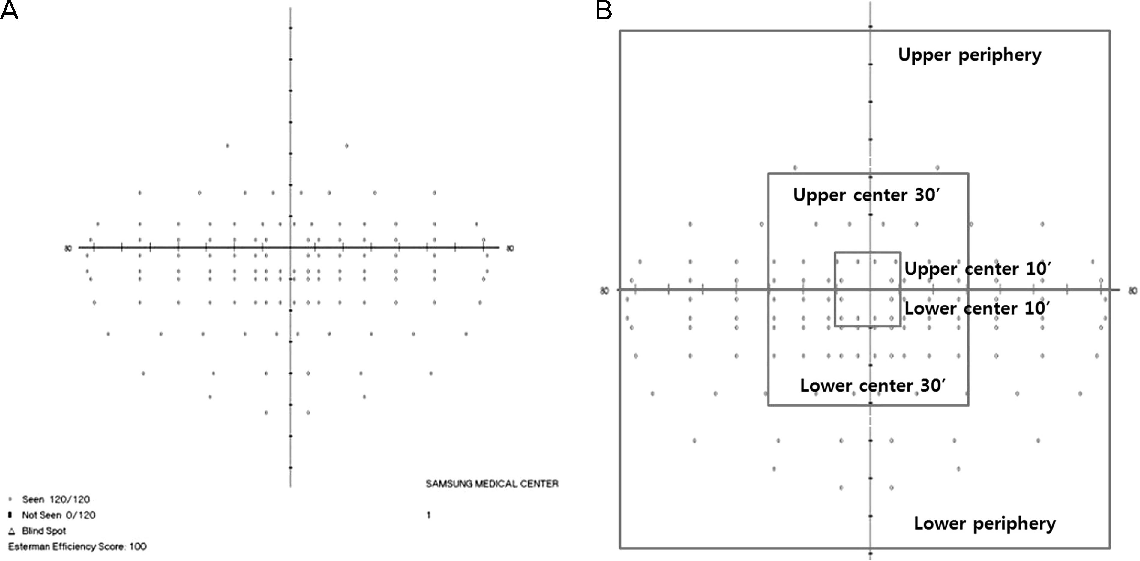
Figure 2.
The average efficient score of each cluster in binocular Esterman visual field Esterman efficient score (%) = (number of point which patients can see) / (total number of point in each cluster) × 100.
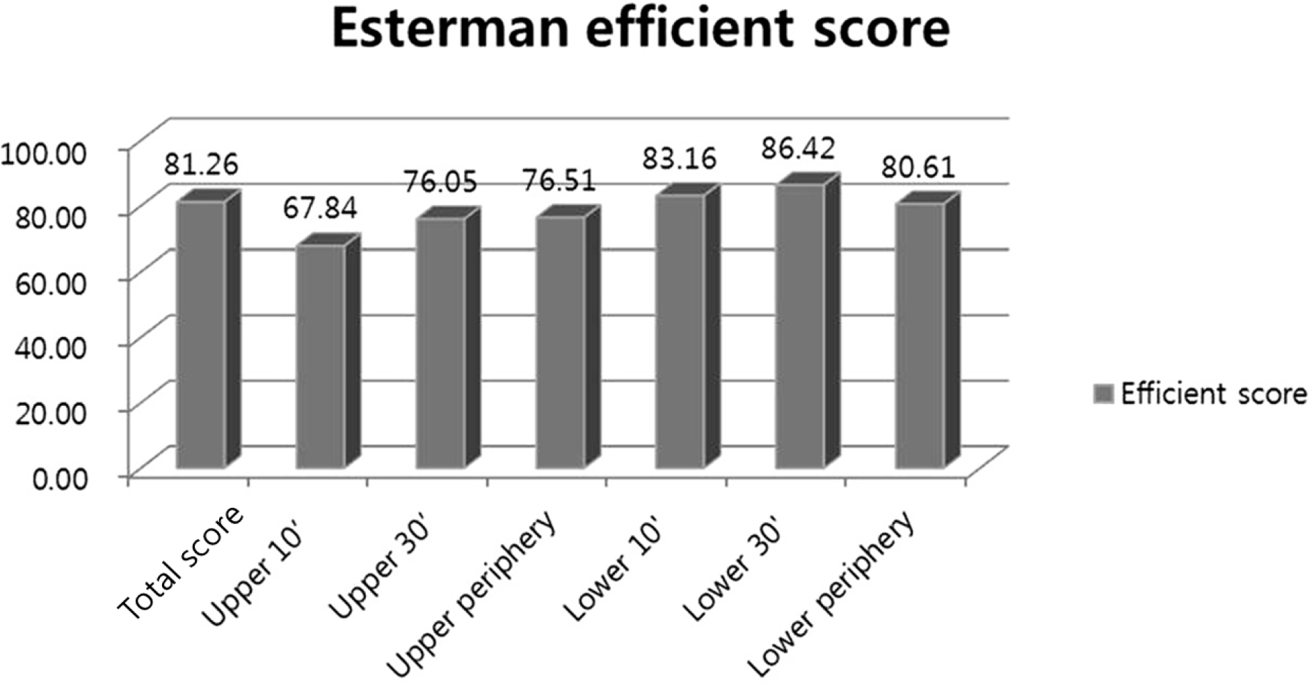
Figure 3.
The average score of each subscale in Visual Function Questionnaire (VFQ). We assessed patients’ vision- specific quality of life by using the Korean version of the 25-item National Eye institute Visual Function Questionnaire (NEI VFQ-25). The VFQ is compsed of 12 subscales and a composite score. The figure showes the average score of each subscale and composition score.
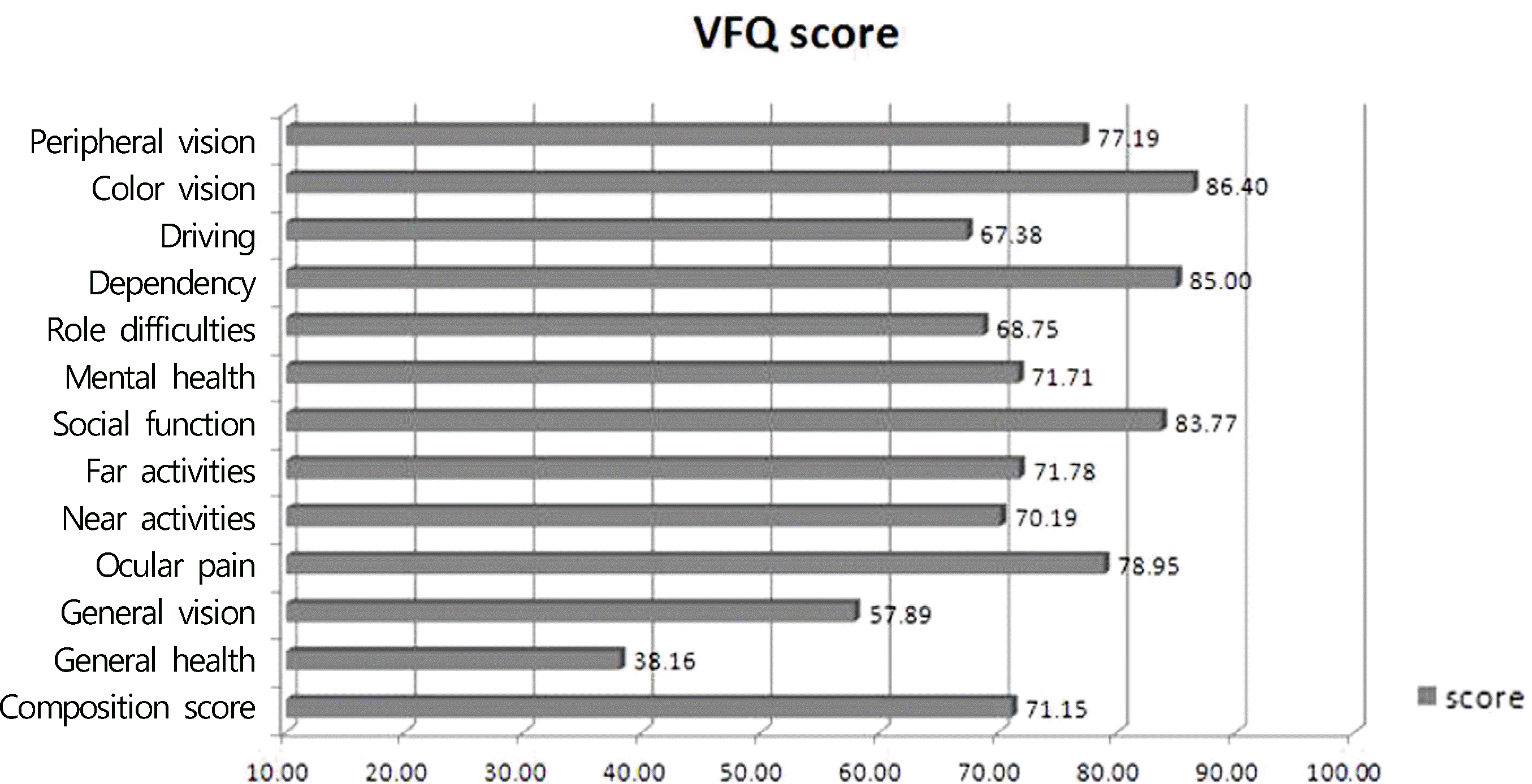
Table 1.
Demographics of the study subjects
Table 2.
The results of correlation coefficients between all clusters of binocular Esterman visual field test and the NEI VFQ-25 (25-item National Eye institute Visual Function Questionnaire)
| Correlation coefficients (n = 60) | Esterman total score | Upper 10’ | Upper 30’ | Upperperiphery | Lower 10’ | Lower 30’ | Lowerperiphery |
|---|---|---|---|---|---|---|---|
| Composition score | 0.40 | 0.08 | 0.23 | 0.44* | 0.28* | 0.22 | 0.40* |
| General health | 0.40* | 0.23 | 0.13 | 0.05 | 0.05 | 0.12 | 0.01 |
| General vision | 0.43 | 0.05 | 0.11 | 0.30* | 0.42* | 0.33* | 0.35* |
| Ocular Pain | 0.01 | 0.02 | 0.60 | 0.09 | 0.08 | 0.03 | 0.04 |
| Near activities | 0.35* | 0.19 | 0.29* | 0.34* | 0.27 | 0.19 | 0.33* |
| Distance activities | 0.37* | 0.11 | 0.24 | 0.40* | 0.28* | 0.21 | 0.38* |
| Social function | 0.29* | 0.10 | 0.18 | 0.32 | 0.32* | 0.25 | 0.29* |
| Mental health | 0.23 | 0.11 | 0.18 | 0.31 | 0.28* | 0.13 | 0.23 |
| Role difficulties | 0.27 | 0.02 | 0.14 | 0.42* | 0.12 | 0.11 | 0.34* |
| Dependency | 0.28* | 0.13 | 0.25 | 0.41* | 0.18 | 0.16 | 0.34* |
| Driving | 0.19 | 0.21 | 0.28 | 0.34* | 0.15 | 0.07 | 0.12 |
| Color vision | 0.29* | 0.13 | 0.28 | 0.40* | 0.20 | 0.23 | 0.36* |
| Peripheral vision | 0.32* | 0.05 | 0.21 | 0.34* | 0.20 | 0.23 | 0.34* |
Table 3.
The results of correlation coefficients between mean deviation of Humphrey Visual Field Analyzer and the NEI VFQ-25 (25-item National Eye institute Visual Function Questionnaire)
| Correlation coefficients (n = 60) | MD (dB) in the better eye | MD (dB) in the worse eye |
|---|---|---|
| Composition score | 0.22 | 0.32* |
| General health | 0.02 | 0.05 |
| General vision | 0.14 | 0.27* |
| Ocular Pain | 0.06 | 0.26 |
| Near activities | 0.17 | 0.33* |
| Distance activities | 0.32 | 0.44* |
| Social function | 0.25 | 0.40* |
| Mental health | 0.23 | 0.14 |
| Role difficulties | 0.07 | 0.23 |
| Dependency | 0.22 | 0.13 |
| Driving | 0.09 | 0.17 |
| Color vision | 0.18 | 0.40* |
| Peripheral vision | 0.14 | 0.32* |
 XML Download