Abstract
Purpose
To report a case of medial canthal tendon fibroma, a rarely observed tumor at the eye or ocular adnexa.
Case summary
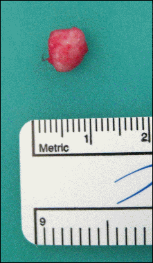
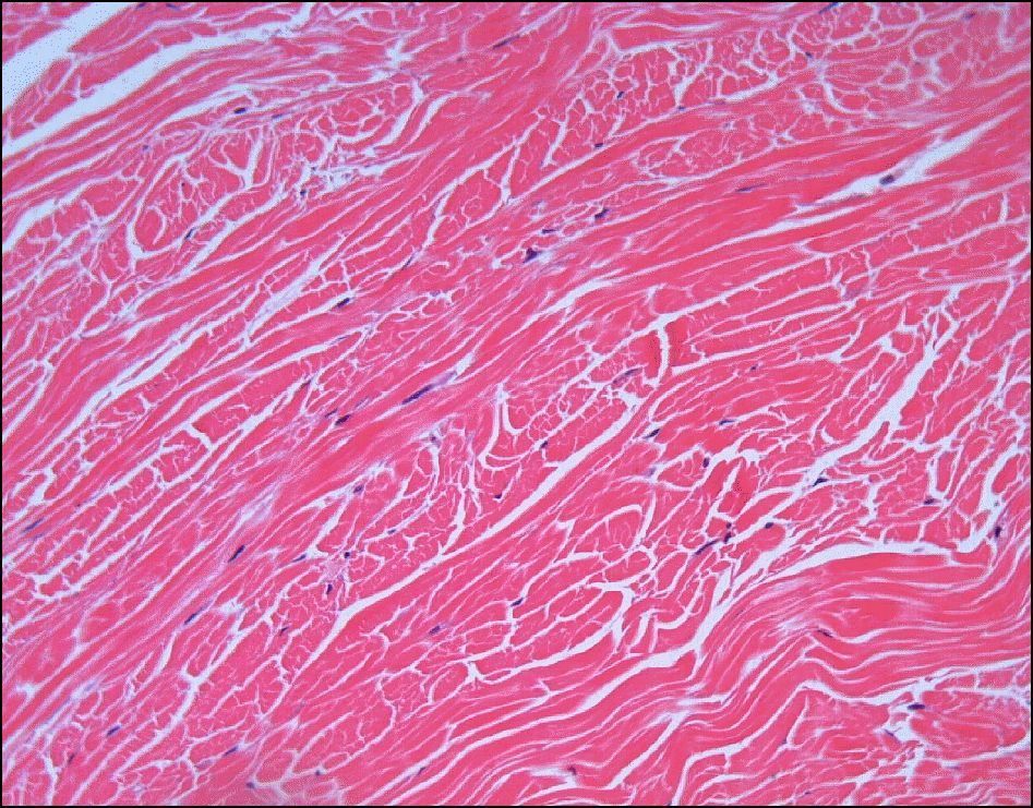
A 47-year-old female visited our clinic with a two-year history of a hard mass in the medial canthal region. On examination, a 7 × 5 mm2 sized, hard and unmovable subcutaneous mass was palpated. The mass was slowly enlarging and the patient had no symptoms including tearing or pain. To confirm the diagnosis, a total excision of the mass was performed under local anesthesia. The tumor was a well-demarcated, 7 × 5 × 2 mm3 sized, white oval mass. The histopathologic examination of the specimen revealed dense collagen bundles with scattered fibroblasts. Based on these findings, the lesion was diagnosed as a fibroma. Although rare, fibromas should be included in the differential diagnosis of medial canthal tumors.
Go to : 
References
1. Herschorn BJ, Jakobiec FA, Hornblass A, et al. Epibulbar subabdominal fibroma. A tumor possibly arising from Tenon's capsule. Ophthalmology. 1983; 90:1490–4.
4. Schutz JS, Rabkin MD, Schutz S. Fibromatous tumor (desmoid type) of the orbit. Arch Ophthalmol. 1979; 97:703–4.

5. Jakobiec FA, Sacks E, Lisman RL, Krebs W. Epibulbar fibroma of the conjunctival substantia propria. Arch of Ophthalmol. 1988; 106:661–4.

6. Choi HT, Ahn M, Moon WS, You IC. A case of tarsal fibroma. J Korean Ophthalmol Soc. 2011; 52:246–9.

7. Kohl SK, Persidsky I, Gigantelli JW. Tendon sheath fibroma of the medial canthus. Ophthal Plast Reconstr Surg. 2007; 23:341–2.

8. Joung Lee MJ, Khwarg SI. Fibroma of the medial canthal area: Aa case report. Ophthal Plast Reconstr Surg. 2011; 27:e21–3.
10. Pulitzer DR, Martin PC, Reed RJ. Fibroma of tendon sheath. A clinicopathologic study of 32 cases. Am J Surg Pathol. 1989; 13:472–9.
11. Yanoff M, Sassani JW. Ocular pathology. 6th ed.China: Elsevier;2009. 188:550–4.
12. Silva P, Bruce IA, Malik T, et al. Nodular fasciitis of the head and neck. J Laryngol Otol. 2005; 119:8–11.

13. Hasegawa T, Matsuno Y, Shimoda T, et al. Extrathoracic solitary fibrous tumors: their histological variability and potentially abdominal behavior. Hum Pathol. 1999; 30:1464–73.
14. Gigantelli JW, Kincaid MC, Soparkar CN, et al. Orbital solitary abdominal tumor: radiographic and histopathologic correlations. Ophthal Plast Reconstr Surg. 2001; 17:207–14.
15. Koeda S, Nagasaka H, Kumamoto H, Kawamura H. Extra-abdominal fibromatosis of the cheek: report of a case. J Oral Maxillofac Surg. 2005; 63:1222–6.

16. O'Connell JX. Pathology of the synovium. Am J Clin Pathol. 2000; 114:773–84.
17. Fitzpatrick TB, Wolff K. Fitzpatrick's dermatology in general abdominal. 7th ed.New York: McGraw-Hill, v. 1.;2008. p. 553–4.
Go to : 
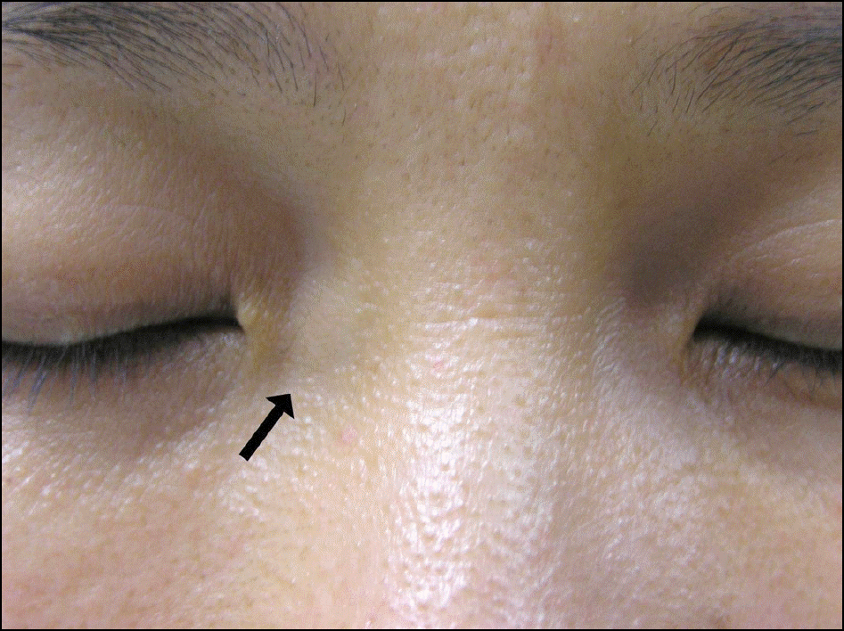 | Figure 1.The subcutaneous mass (black arrow) is visible on the medial canthal area. It was rock-hard and unmovable. |
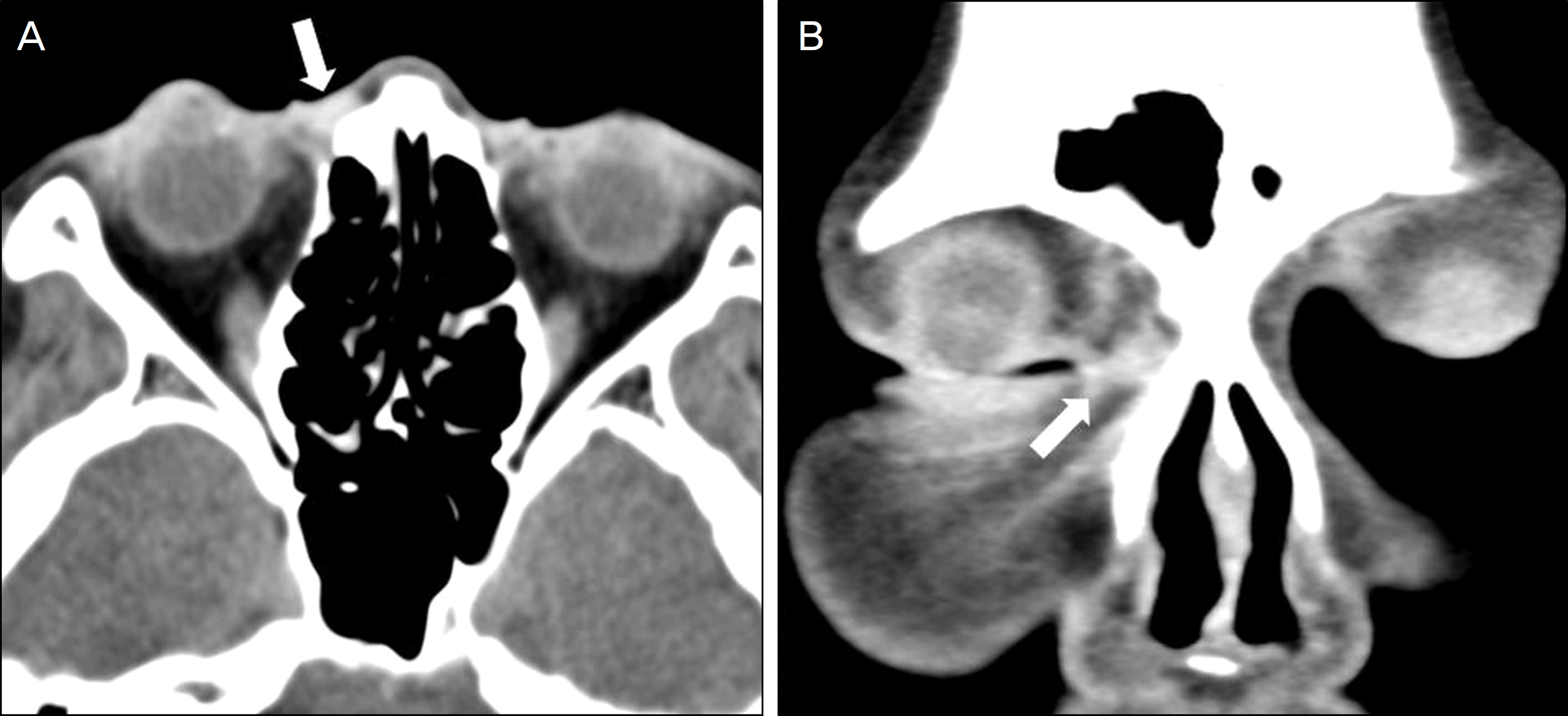 | Figure 2.Axial (A) and coronal (B) orbital CT show a 7 × 5 mm2 sized, ill demarcated, focally enhancing preseptal lesion near the right medial canthus (white arrows). |
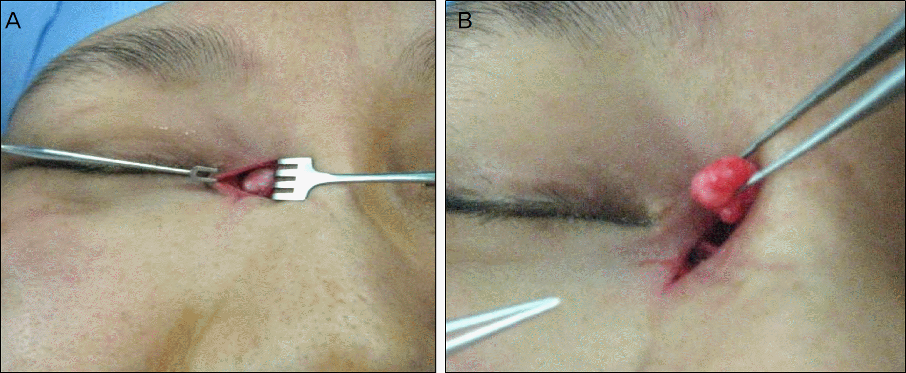 | Figure 3.The photographs show(A) well demarcated mass at the medial canthal area. (B) The floor of mass was firm-ly attached to periosteum. |


 PDF
PDF ePub
ePub Citation
 Print
Print


 XML Download