

 PDF
PDF ePub
ePub Citation
 Print
Print


Abstract
Purpose
In this study we evaluated the changes in the corneal endothelial cells before and after the operation among myopes in the M-LASEK group, on whom 0.02% mitomycin C (MMC) was used and in the LASEK group, on whom MMC was not used.
Methods
The corneal endothelial cell analysis was performed in 104 eyes of 57 subjects in the LASEK group and in 86 eyes of 48 subjects in the M-LASEK group before the operation, and 3 months and 12 months postoperatively.
Results
There were no statistically significant differences in the corneal endothelial cell density (CD), the cell area coefficient of variance (CV), and hexagonal cell rate (6A) between the 2 groups before the operation, and 3 months and 12 months postoperatively (p> 0.05). In the LASEK group, there were no statistically significant differences (p> 0.05) in CD when the numerical values before the operation and 3 months and 12 months after the operation were compared, but there were statistically significant differences in CV and 6A when comparing before the operation and 12 months postoperatively (p= 0.001, p = 0.034, respectively). In the M- LASEK group, there was a 2.8% statistically significant decrease (p= 0.004) in CD when the numerical values before the operation and 3 months after the operation were compared, but there were no statistically significant difference (p> 0.05) when the numerical values before the operation and 12 months after the operation were compared. In addition, there were no statistically significant differences (p> 0.05) in CV and 6A when the numerical values before the operation and 3 months and 12 months after the operation were compared.
Go to : 
References
1. Camellin M. Laser epithelial keratomileusis for myopia. J Refract Surg. 2003; 19:666–70.

2. Taneri S, Zieske JD, Azar DT. Evolution, techniques, clinical outcomes, and pathophysiology of LASEK: Review of the literature. Surv Ophthalmol. 2004; 49:576–602.
3. Chang SW. Early corneal edema following topical application of mitomycin-C. J Cataract Refract Surg. 2004; 30:1742–50.
4. Xu H, Liu S, Xia X. . Mitomycin C reduces haze formation in rabbits after excimer laser photorefractive keratectomy. J Refract Surg. 2001; 17:342–9.
5. Majmudar PA, Forstot SL, Dennis RF. . Topical mitomycin-C for subepithelial fibrosis after refractive corneal surgery. Ophthalmology. 2000; 107:89–94.
6. Carones F, Vigo L, Scandola E, Vacchini L. Evaluation of the prophylactic use of mitomycin-C to inhibit haze formation after photorefractive keratectomy. J cataract Refract Surg. 2002; 28:2088–95.
7. Lai YH, Wang HZ, Lin CP, Chang SJ. Mitomycin C alters corneal stromal wound healing and corneal haze in rabbits after argon-fluoride excimer laser photorefractive keratectomy. J Ocul Pharmacol Ther. 2004; 20:129–38.
8. Hashemi H, Taheri SM, Fotouhi A, Kheiltash A. Evaluation of the prophylactic use of mitomycin C to inhibit haze formation after photorefractive keratectomy in high myopia: a prospective clinical study. BMC Ophthalmol. 2004; 4:12.
9. Gambato C, Ghirlando A, Moretto E. . Mitomycin C modulation of corneal wound healing after photorefractive keratectomy in highly myopic eyes. Ophthalmology. 2005; 112:208–18. discussion 219.
10. Pfister RR. Permanent corneal edema resulting from the treatment of PTK corneal haze with mitomycin: a case report. Cornea. 2004; 23:744–7.
11. Kim TI, Tchah H, Lee SA. . Apoptosis in keratocytes caused by mitomycin C. Invest Ophthalmol Vis Sci. 2003; 44:1912–7.
12. Kim TI, Pak JH, Lee SY, Tchah H. Mitomycin C-induced reduction of keratocytes and fibroblasts after photorefractive keratectomy. Invest Ophthalmol Vis Sci. 2004; 45:2978–84.
13. Winkler von Mohrenfels C, Reischl U, Lohmann CP. Corneal haze after photorefractive keratectomy for myopia: role of collagen IV mRNA typing as a predictor of haze. J Cataract Refract Surg. 2002; 28:1446–51.
14. Kim TI, Tchah H, Cho EH, Kook MS. Evaluation for safety of cultured corneal fibroblasts with cotreatment of alcohol and mitomycin C. Invest Ophthalmol Vis Sci. 2004; 45:86–92.
15. Zhou J, Lu S, Dai J. . Short-term corneal endothelial changes after laser-assisted subepithelial keratectomy. J Int Med Res. 2010; 38:1484–90.
16. Jung YH, Chung SK. Corneal endothelial changes after laser-assisted subepithelial keratomileusis. J Korean Ophthalmol Soc. 2013; 54:33–37.
17. Zhao LQ, Wei RL, Ma XY, Zhu H. Effect of intraoperative mito- mycin-C on healthy corneal endothelium after laser-assisted subepithelial keratectomy. J Cataract Refract Surg. 2008; 34:1715–9.
18. de Benito-Llopis L, Teus MA, Ortega M. Effect of mitomycin-C on the corneal endothelium during excimer laser surface ablation. J Cataract Refract Surg. 2007; 33:1009–13.
19. Nishida T, Saika S. Cornea and Sclera: anatomy and physiology. Krachmer JH, Mannis MJ, Holland EJ, editors. Cornea. 3rd ed.St. Louis: Mosby;2011. p. 15–6.
20. Park YJ, Lee GJ, Park JJ. . The long-term effects of soft contact lens wear on corneal thickness, curvature and endothelium. J Korean Ophthalmol Soc. 2005; 46:945–53.
21. Stefansson E, Wolbarst ML, Landers MB 3rd. Corneal contact lens and aqueous humor hypoxia in cat. Invest Ophthalmol Vis Sci. 1983; 24:1052–4.
22. Patel SV, Bourne WM. Corneal endothelial cell loss 9 years after excimer laser kearatorefractive surgery. Arch Ophthalmol. 2009; 127:1423–7.
23. Lee JH, Ahn JH, Lew HM, Lee DH. Effect of mitomycin C on rabbit corneal wound healing after excimer laser photorefractive keratectomy. J Korean Ophthalmol Soc. 2003; 44:2876–84.
24. Jeong BJ, Kim HH, Park YJ. . Effect of mitomycin C to inhibit corneal haze formation after photorefractive keratectomy for high myopia. J Korean Ophthalmol Soc. 2006; 47:725–34.
25. Pallikaris IG, Síganos DS. Laser in situ keratomileusis to treat myopia: early experience. J Cataract Refract Surg. 1997; 23:39–49.
Go to :
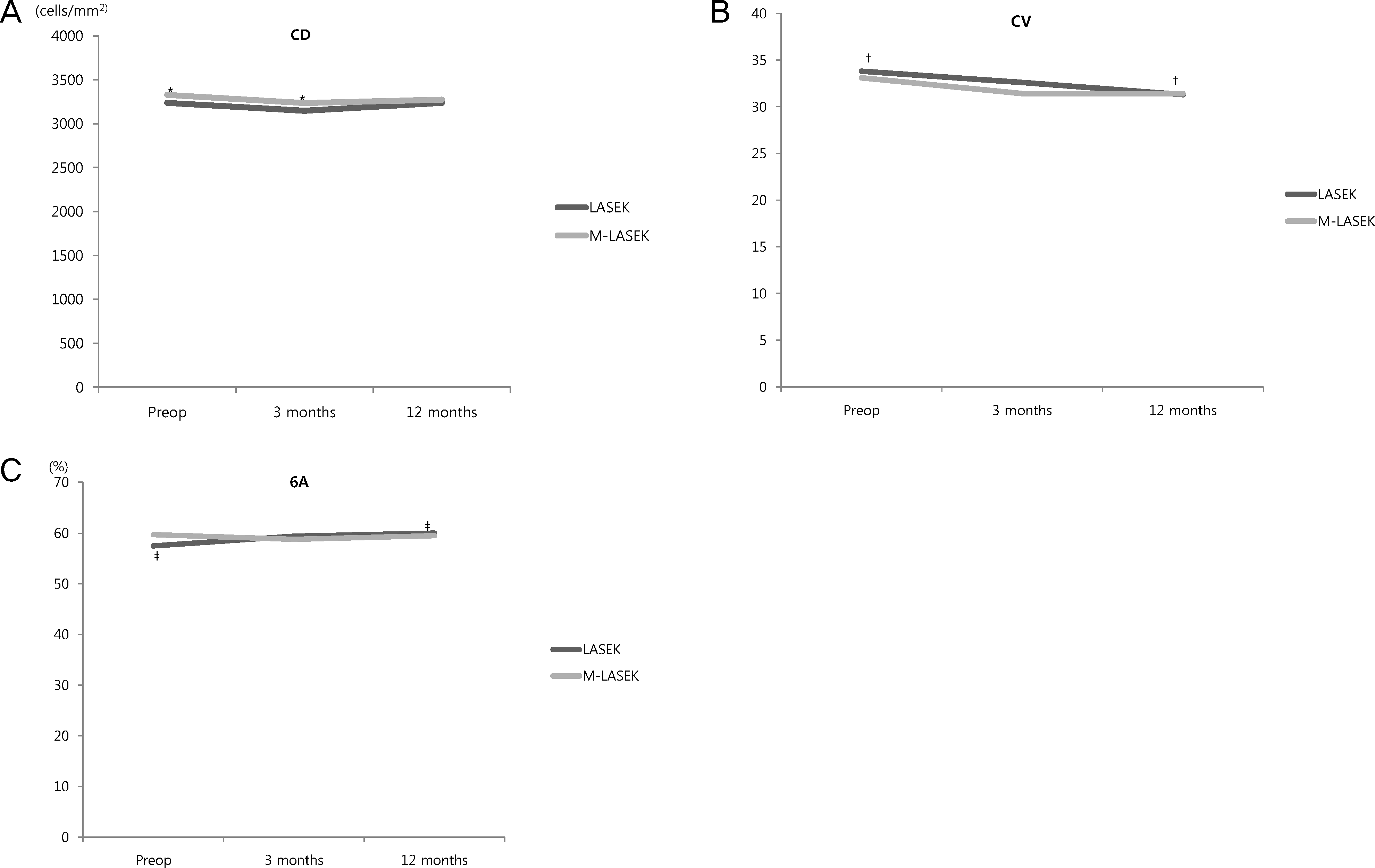 | Figure 1.Corneal endothelial changes after LASEK and M-LASEK. Paired t-test *p = 0.004; †p = 0.001; ‡p = 0.034. CD = endothelial cell density; CV = coefficient of variation of cell area; 6A = hexagonality. |
Table 1.
Preoperative demographics of patients in LASEK group and M-LASEK group
| LASEK | M*-LASEK | p-value† | |
|---|---|---|---|
| Age (years) | 28.6 ± 7.8 (19∼50) | 26.9 ± 6.9 (18∼43) | 0.260 |
| Male : Female (eyes) | 19 : 85 | 21 : 65 | 0.654 |
| Preoperative SE (D) | −4.04 ± 1.20 (-1.38∼-6.38) | −7.09 ± 1.15 (-5.0∼-9.5) | <0.001 |
| Corneal thickness (μm) | 543 ± 35 (464∼651) | 541 ± 31 (482∼604) | 0.727 |
| Ablation thickness (μm) | 87 ± 16 (40∼112) | 129 ± 12 (104∼151) | <0.001 |
| Total (eyes) | 104 | 86 |
Table 2.
Preoperative and postoperative data for the cell density (CD), the coefficient of variation (CV), the hexagonality (6A) in two groups
| CD (cells/mm2) | CV | 6A (%) | |
|---|---|---|---|
| Preop | |||
| LASEK (n = 104) | 3237 ± 322 | 33.8 ± 5.6 | 57.5 ± 9.7 |
| M-LASEK (n = 86) | 3328 ± 304 | 33.1 ± 7.8 | 59.7 ± 11.6 |
| p-value* | 0.081 | 0.405 | 0.187 |
| Postop 3 months | |||
| LASEK (n = 48) | 3147 ± 353 | 32.6 ± 5.4 | 59.4 ± 5.4 |
| M-LASEK (n = 60) | 3236 ± 315 | 31.4 ± 5.1 | 58.8 ± 10.7 |
| p-value* | 0.172 | 0.232 | 0.745 |
| Postop 12 months | |||
| LASEK (n = 104) | 3238 ± 320 | 31.3 ± 5.2 | 60.0 ± 10.0 |
| M-LASEK (n = 86) | 3274 ± 292 | 31.4 ± 5.3 | 59.5 ± 10.9 |
| p-value* | 0.432 | 0.891 | 0.729 |
Table 3.
Changes in corneal endothelium indices after LASEK
| Time | CD (cells/mm2) | p-value* | CV | p-value* | 6A (%) | p-value* |
|---|---|---|---|---|---|---|
| Preop (n = 104) | 3237 ± 322 | − | 33.8 ± 5.6 | − | 57.5 ± 9.7 | − |
| Postop | ||||||
| 3 months (n = 48) | 3147 ± 353 | 0.083 | 32.6 ± 5.4 | 0.122 | 59.4 ± 5.4 | 0.203 |
| 12 months (n = 104) | 3238 ± 320 | 0.628 | 31.3 ± 5.2 | 0.001 | 60.0 ± 10.0 | 0.034 |
Table 4.
Changes in corneal endothelium indices after M-LASEK
| Time | CD (cells/mm2) | p-value* | CV | p-value* | 6A (%) | p-value* |
|---|---|---|---|---|---|---|
| Preop (n = 86) | 3328 ± 304 | − | 33.1 ± 7.8 | − | 59.7 ± 11.6 | − |
| Postop | ||||||
| 3 months (n = 60) | 3236 ± 315 | 0.004 | 31.4 ± 5.1 | 0.147 | 58.8 ± 10.7 | 0.682 |
| 12 months (n = 86) | 3274 ± 292 | 0.055 | 31.4 ± 5.3 | 0.057 | 59.5 ± 10.9 | 0.905 |
 XML Download