

 PDF
PDF ePub
ePub Citation
 Print
Print


Abstract
Purpose
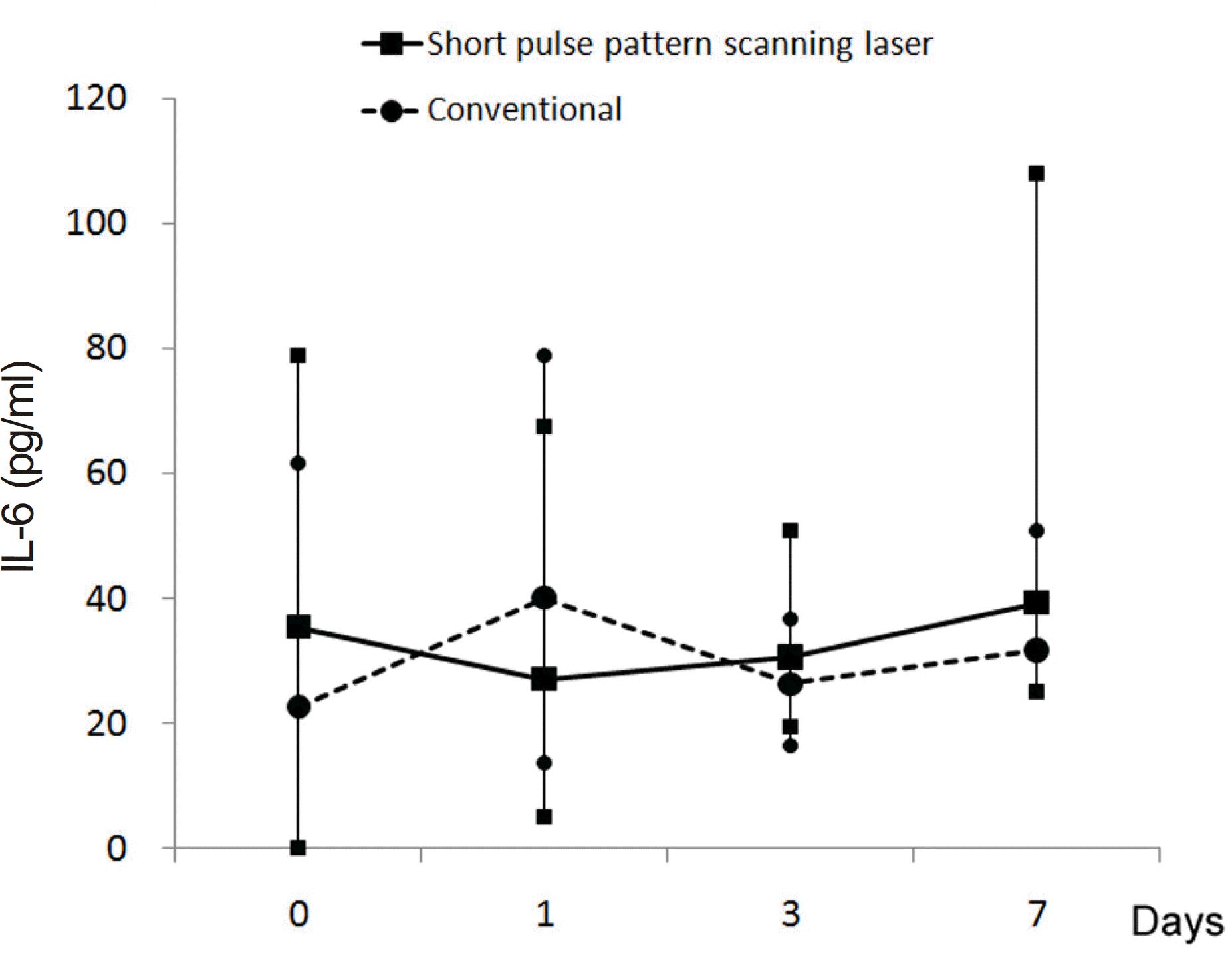
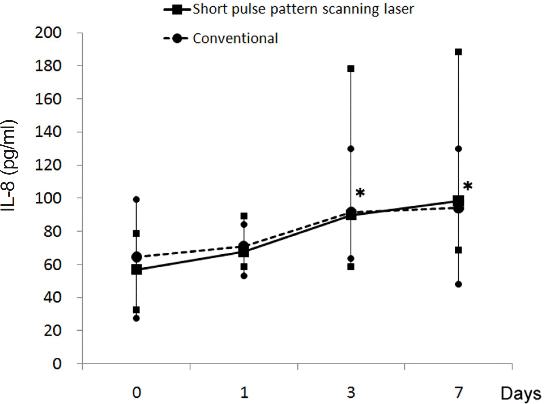
To compare concentration of cytokines in the vitreous of rabbit eyes after photocoagulation using a short-pulse pattern scanning laser (PASCAL) or a conventional laser.
Methods
Laser photocoagulation was performed using PASCAL (duration 0.02 seconds) in the right eyes and a conventional laser (duration 0.1 seconds) in the left eyes of 13 pigmented rabbits. To obtain ophthalmoscopically similar mild burns, power was adjusted during the photocoagulation. The rabbits were sacrificed at 1, 3 or 7 days after photocoagulation to investigate histological changes. Levels of interleukins (IL)-1β, IL-6, IL-8 and tumor necrosis factor (TNF)-α in the vitreous humors of ten rabbits were measured using enzyme-linked immunosorbent assay before treatment and at 1, 3 and 7 days after photocoagulation.
References
1. Techniques for scatter and local photocoagulation treatment of abdominal retinopathy: Early Treatment Diabetic Retinopathy Study Report no. 3. The Early Treatment Diabetic Retinopathy Study Research Group. Int Ophthalmol Clin. 1987; 27:254–64.
2. McDonald HR, Schatz H. Visual loss following panretinal photo-coagulation for proliferative diabetic retinopathy. Ophthalmology. 1985; 92:388–93.

3. McDonald HR, Schatz H. Macular edema following panretinal photocoagulation. Retina. 1985; 5:5–10.
4. Shimura M, Yasuda K, Nakazawa T, Tamai M. Visual dysfunction after panretinal photocoagulation in patients with severe diabetic retinopathy and good vision. Am J Ophthalmol. 2005; 140:8–15.
5. Er H, Doganay S, Turkoz Y, et al. The levels of cytokines and nitric oxide in rabbit vitreous humor after retinal laser photocoagulation. Ophthalmic Surg Lasers. 2000; 31:479–83.
6. Shimura M, Yasuda K, Nakazawa T, et al. Panretinal photo-coagulation induces proinflammatory cytokines and macular thickening in high-risk proliferative diabetic retinopathy. Graefes Arch Clin Exp Ophthalmol. 2009; 247:1617–24.
7. Early photocoagulation for diabetic retinopathy. ETDRS report number 9. Early Treatment Diabetic Retinopathy Study Research Group. Ophthalmology. 1991; 98(5 Suppl):766–85.
8. Margolis R, Singh RP, Bhatnagar P, Kaiser PK. Intravitreal abdominal as adjunctive treatment to laser panretinal photo-coagulation for concomitant proliferative diabetic retinopathy and clinically significant macular oedema. Acta Ophthalmol. 2008; 86:105–10.
9. Shimura M, Yasuda K, Shiono T. Posterior abdominal's capsule abdominal of triamcinolone acetonide prevents panretinal photo-coagulation-induced visual dysfunction in patients with severe abdominal retinopathy and good vision. Ophthalmology. 2006; 113:381–7.
10. Blumenkranz MS, Yellachich D, Andersen DE, et al. Semiautomated patterned scanning laser for retinal photocoagulation. Retina. 2006; 26:370–6.
11. Luttrull JK, Musch DC, Spink CA. Subthreshold diode micropulse panretinal photocoagulation for proliferative diabetic retinopathy. Eye (Lond). 2008; 22:607–12.
12. Al-Hussainy S, Dodson PM, Gibson JM. Pain response and fol-low-up of patients undergoing panretinal laser photocoagulation with reduced exposure times. Eye (Lond). 2008; 22:96–9.
13. Sanghvi C, McLauchlan R, Delgado C, et al. Initial experience with the Pascal photocoagulator: a pilot study of 75 procedures. Br J Ophthalmol. 2008; 92:1061–4.
14. Bhattacherjee P, Henderson B. Inflammatory responses to abdominally injected interleukin 1. Curr Eye Res. 1987; 6:929–34.
15. Palexas GN, Sussman G, Welsh NH. Ocular and systemic abdominal of IL-1 beta and tumour necrosis factor in a patient with ocular inflammation. Scand J Immunol Suppl. 1992; 11:173–5.
16. Jirik FR, Podor TJ, Hirano T, et al. Bacterial lipopolysaccharide and inflammatory mediators augment IL-6 secretion by human abdominal cells. J Immunol. 1989; 142:144–7.
17. Funatsu H, Yamashita H, Ikeda T, et al. Vitreous levels of inter-leukin-6 and vascular endothelial growth factor are related to abdominal macular edema. Ophthalmology. 2003; 110:1690–6.
18. Photocoagulation treatment of proliferative diabetic retinopathy. Clinical application of Diabetic Retinopathy Study (DRS) abdominal, DRS Report Number 8. The Diabetic Retinopathy Study Research Group. Ophthalmology. 1981; 88:583–600.
19. Zacks DN, Johnson MW. Combined intravitreal injection of triamcinolone acetonide and panretinal photocoagulation for abdominal diabetic macular edema and proliferative diabetic retinopathy. Retina. 2005; 25:135–40.
20. Margolis R, Singh RP, Bhatnagar P, Kaiser PK. Intravitreal abdominal as adjunctive treatment to laser panretinal photo-coagulation for concomitant proliferative diabetic retinopathy and clinically significant macular oedema. Acta Ophthalmol. 2008; 86:105–10.
21. Jain A, Blumenkranz MS, Paulus Y, et al. Effect of pulse duration on size and character of the lesion in retinal photocoagulation. Arch Ophthalmol. 2008; 126:78–85.
Figure 1.
Ophthalmoscopic findings after photocoagulation using pattern scanning laser (A: day 0; C: day 1; E: day 3; G: day 7) and conventional laser (B: day 0; D: day 1; F: day 3; H: day 7).
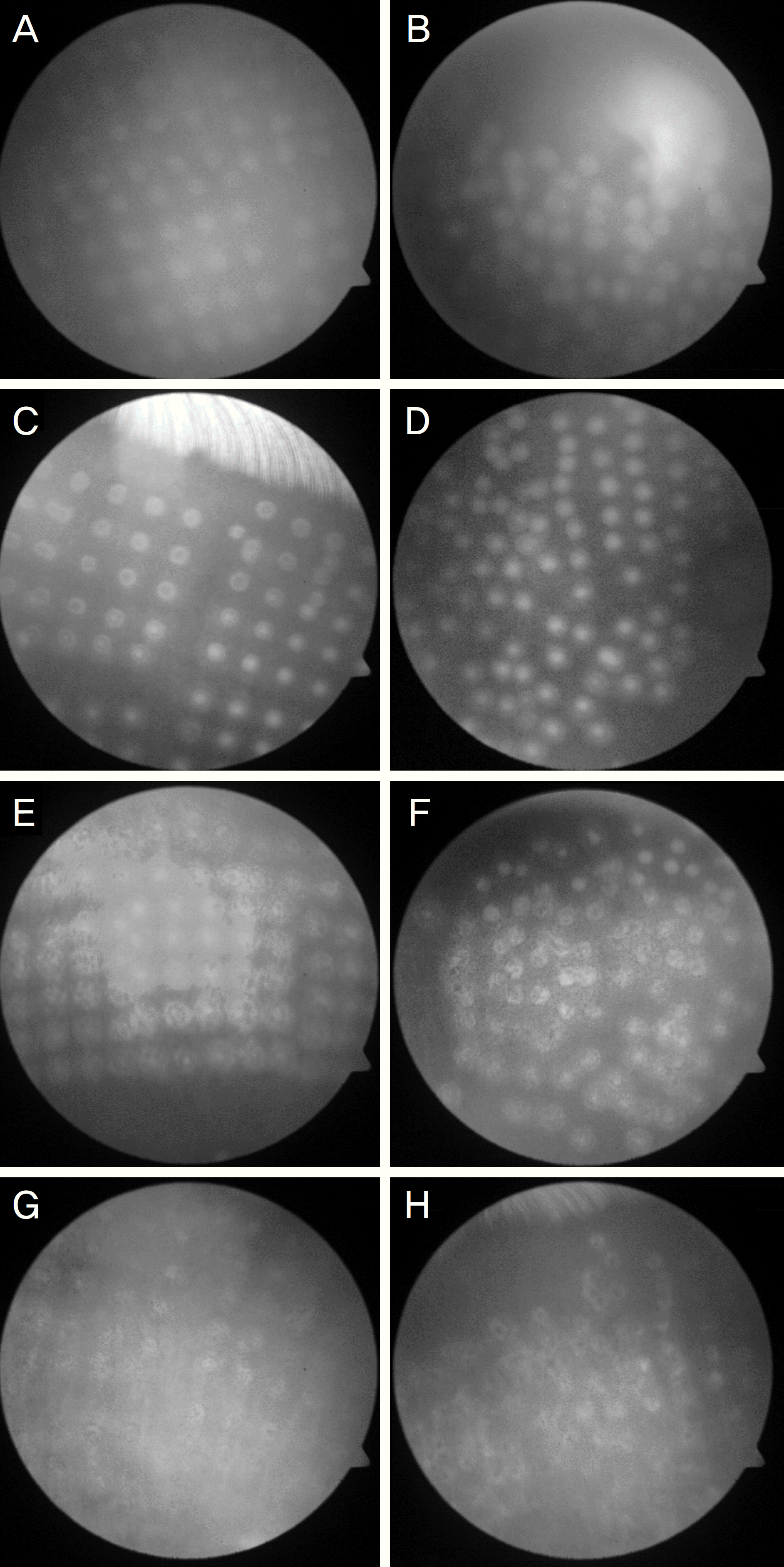
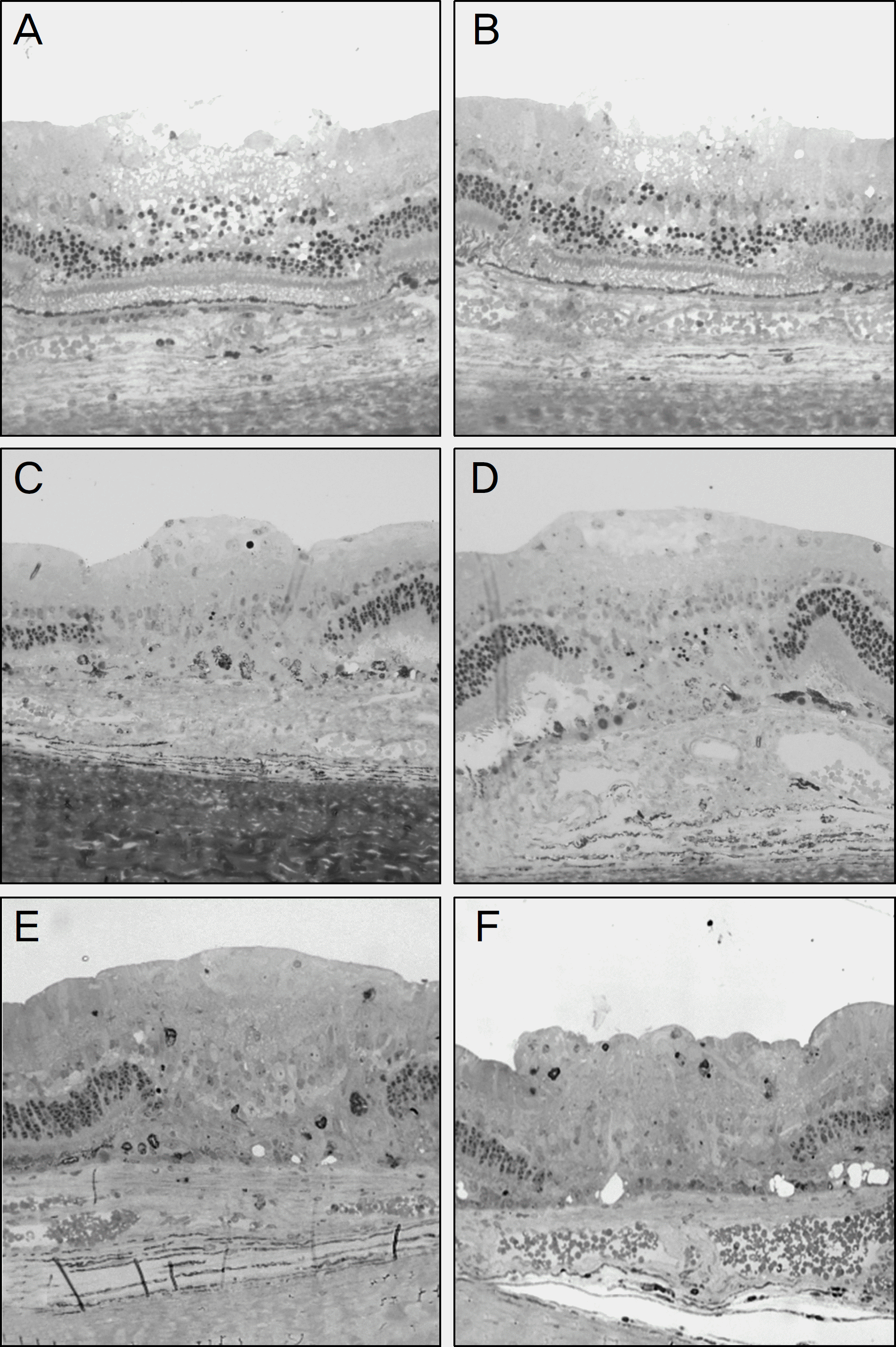
Figure 2.
Microscopic findings after photocoagulation using pattern scanning laser (A: day 1; C: day 3; E: day 7) and conventional laser (B: day 1; D: day 3; F: day 7).
 XML Download