

 PDF
PDF ePub
ePub Citation
 Print
Print


Abstract
Purpose
To report on the clinical manifestations, species and treatments of patients with chronic canaliculitis.
Methods
From August 2003 to February 2012, 77 eyes of 77 patients who were diagnosed with chronic canaliculitis at our hospital were retrospectively analyzed.
Results
The mean period from the onset of symptoms to diagnosis was 4.7 months. The most common systemic disease associated with chronic canaliculitis was diabetes (18 eyes, 23%), and 13 eyes (17%) were related to punctual plug insertion. Main symptoms consisted of epiphora with discharge and pouting punctum. In the culture results of 55 eyes, streptococci, staphylococci, and actinomyces among other bacteria were identified. Seventy-two eyes (94%) were cured with one-snip punctoplasty with curettage.
Conclusions
Chronic canaliculitis is rare, and the clinical aspect can be obscured by chronic conjunctivitis, thus the diagnosis is often delayed. In patients who have systemic diseases such as diabetes or past history of punctual plug insertion, chronic canaliculitis should be differentiated by observing the punctum more closely. If the diagnosis is accurate at the time, chronic canaliculitis could be easily cured by a relatively simple procedure such as one-snip punctoplasty with curettage.
References
1. Kim SD, Koh SI, Kim JD. Diagnosis and therapy of canaliculitis. J Korean Ophthalmol Soc. 1998; 39:2207–10.
2. Hong JW, Lee TS. Two cases of chronic canaliculitis. J Korean Ophthalmol Soc. 1990; 31:1096–102.
3. Briscoe D, Edelstein E, Zacharopoulos I. . Actinomyces canaliculitis: diagnosis of a masquerading disease. Arch Clin Exp Ophthalmol. 2004; 242:682–6.

4. Freedman JR, Markert MS, Cohen AJ. Primary and secondary lacrimal canaliculitis: a review of literature. Surv Ophthalmol. 2011; 56:336–47.
5. Chen SX, Lee GA. SmartPlug in the management of severe dry eye syndrome. Cornea. 2007; 26:534–8.
6. Varma D, Chang B, Musaad S. A case series on chronic canaliculitis. Orbit. 2005; 24:11–4.
7. Baldursdóttir E, Sigurdsson H, Jónasson L, Gottfredsson M. Actinomycotic canaliculitis: resolution following surgery and short topical antibiotic treatment. Acta Ophthalmol. 2010; 88:367–70.
8. Vécsei VP, Huber-Spitzy V, Arocker-Mettinger E, Steinkogler FJ. Canaliculitis: Difficulties in diagnosis, differential diagnosis and comparison between conservative and surgical treatment. Ophthalmologica. 1994; 208:314–7.
9. Zaldivar RA, Bradley EA. Primary canaliculitis. Ophthal Plast Reconstr Surg. 2009; 25:481–4.
10. Lin SC, Kao SC, Tsai CC. . Clinical characteristics and factors associated the outcome of lacrimal canaliculitis. Acta Ophthalmol. 2011; 89:759–63.
11. Pavilack MA, Frueh BR. Thorough curettage in the treatment of chronic canaliculitis. Arch Ophthalmol. 1992; 110:200–2.
12. Nelson CC. Complications of Freeman plugs. Arch Ophthalmol. 1991; 109:923–4.
13. Jang JH, Chae JK, Kim BJ, Lee HB. Cases of complications after the use of punctal plugs. J Korean Ophthalmol Soc. 2005; 46:547–53.
14. Lim DK, Joo MJ, Kim JH. A case of chronic granulomatous canaliculitis induced by herrick silicone punctal plug. J Korean Ophthalmol Soc. 2005; 46:384–7.
15. Mohan ER, Kabra S, Udhay P, Madhavan HN. Intracanalicular antibiotics may obviate the need for surgical management of chronic suppurative canaliculitis. Indian J Ophthalmol. 2008; 56:338–40.
16. Ahn SM, Kim HC, Jang JW, Kim SJ. Treatment of the SmartPLUG- related canaliculitis. J Korean Ophthalmol Soc. 2009; 50:1768–73.
17. SmartPlug Study Group. Management of complications after in-sertion of the SmartPlug punctal plug: a study of 28 patients. Ophthalmology. 2006; 113:1859; e1–6.
18. Lee MJ, Choung HK, Kim NJ, Khwarg SI. One-snip punctoplasty and canalicular curettage through the punctum: a minimally invasive surgical procedure for primary canaliculitis. Ophthalmology. 2009; 116:2027–30.e2.
19. Lee MJ, Lee KW, Kim NJ. . Canaliculitis associated with SmartPLUGTM punctal plug insertion: clinical features and management. J Korean Ophthalmol Soc. 2009; 50:821–5.
20. Lee J, Flanagan JC. Complications associated with silicone intra- canalicula plugs. Ophthal Plast Reconstr Surg. 2001; 17:465–9.
21. White WL, Bartley GB, Hawes MJ. . Iatrogenic complications related to the use of Herrick lacrimal plugs. Ophthalmology. 2001; 108:1835–7.
Figure 1.
(A) Pouting punctum, Injection of the conjunctiva could be misdiagnosed as chronic recurrent conjunctivitis. (B) Mucopurulent punctual regurgitation. (C) Peripunctal erythematous swelling. (D) Pyogenic granuloma.
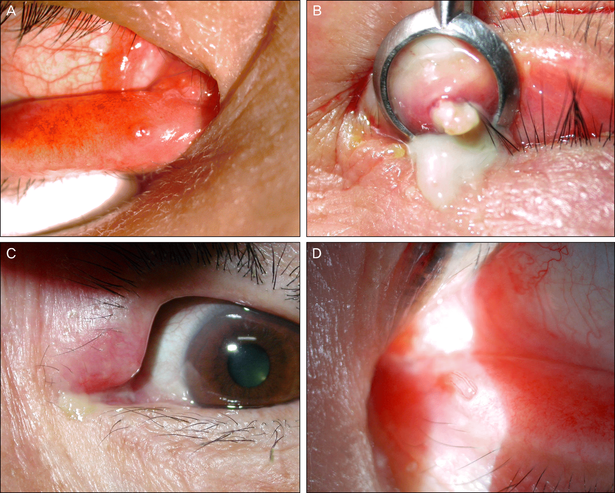
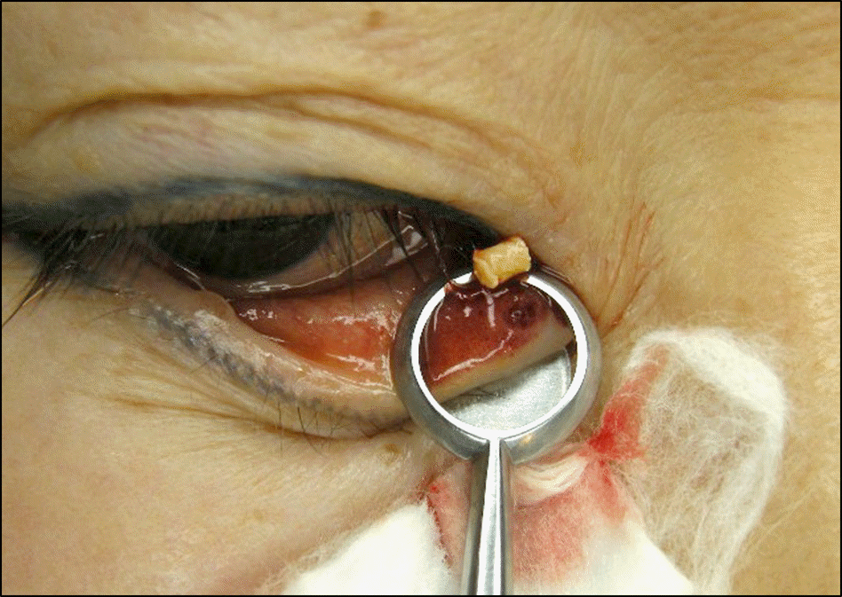
Figure 3.
(A) Freeman-type punctal plugs were eliminated from the left lower punctum. (B) This picture shows the removed Freeman-type punctal plugs. Note the two Freeman-type punctal plugs had been inserted in the same punctum.
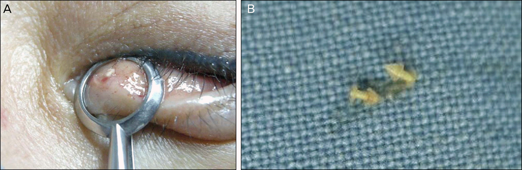
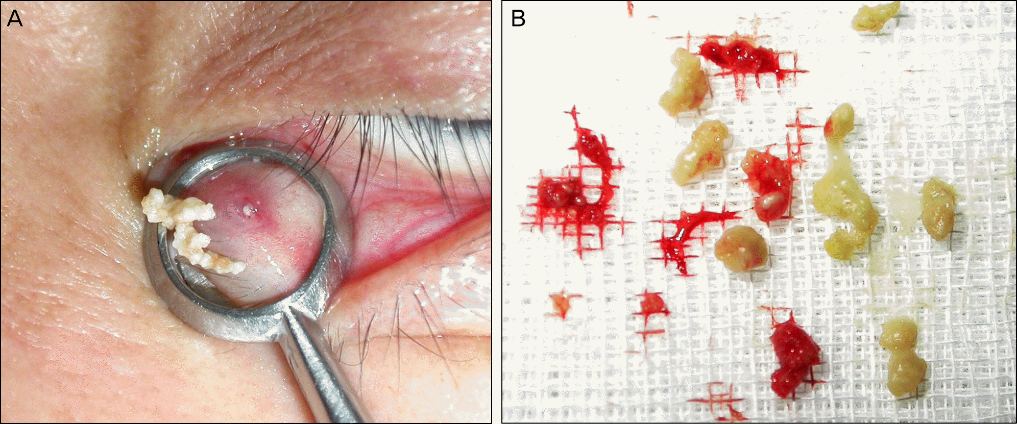
Figure 5.
(A) Photograph showing the punctum of a patient with recurrent canaliculitis. (B) Two weeks after punctoplasty and curettage, the signs of canaliculitis had resolved completely.
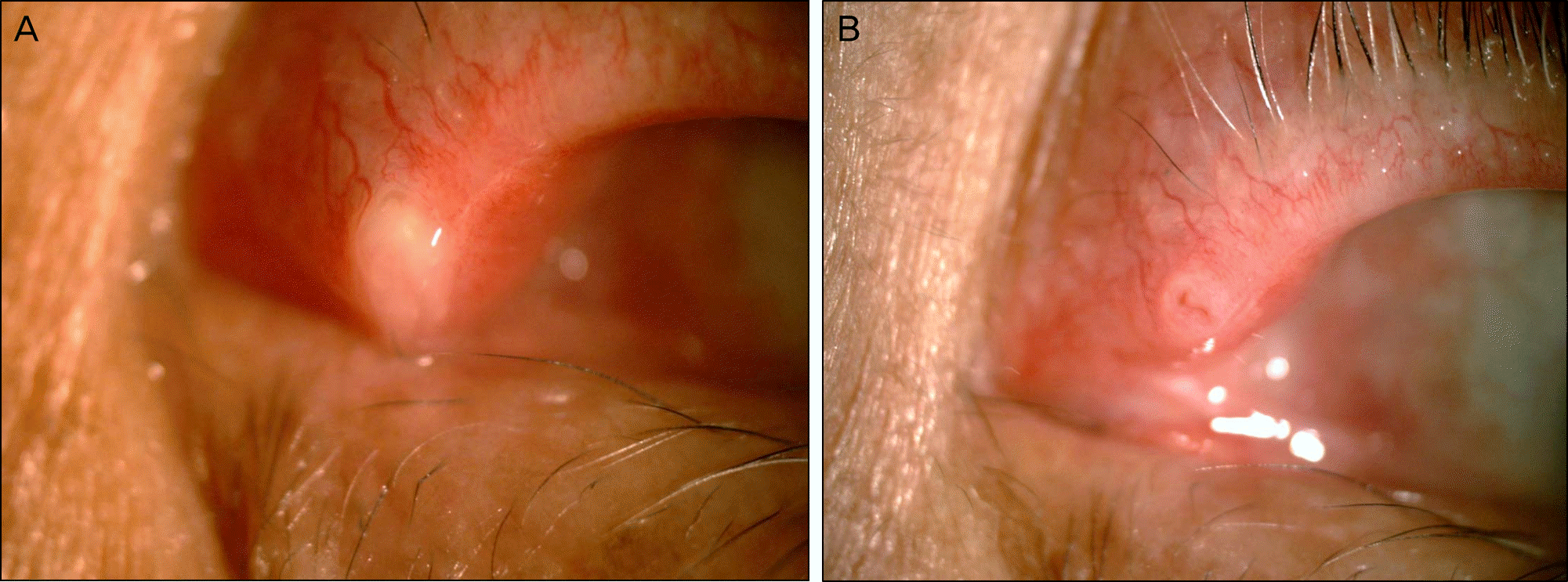
Table 1.
Clinical characteristics, treatment, and outcome for the 77 patients studied
Table 2.
Clinical presentation and treatment of patients with canaliculitis after punctual plug insertion
Table 3.
Microbiologie findings of 55 cultured lacrimal canaliculits
Table 4.
Clinical presentation of the recurred patients
 XML Download