

 PDF
PDF ePub
ePub Citation
 Print
Print


Abstract
Purpose
To report a case of globe perforation and linear retinal tear after periocular acupuncture therapy which resulted in persistent temporal field defect with normal retinal function evidenced by multifocal electroretinogram (MERG).
Case summary
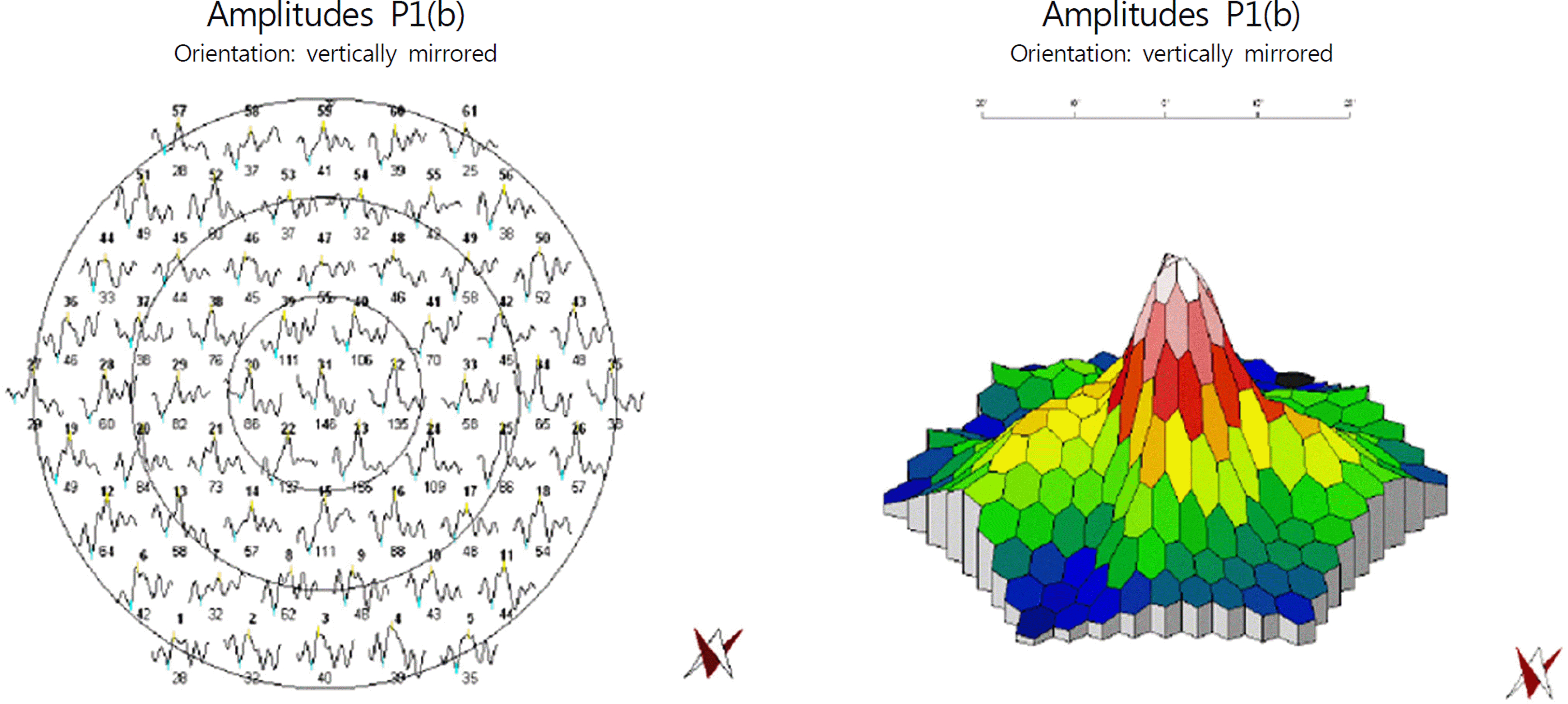
A 42-year-old female presented with decreased visual acuity and pain in her right eye after a periocular acupuncture therapy for blepharospasm. At initial presentation, the best corrected visual acuity (BCVA) was 0.08 in the in-jured eye and the intraocular pressure was 15 mmHg. Ultrasonography showed minimal vitreous hemorrhage and fundus examination revealed a linear retinal tear in the posterior pole sparing the macula. Consequently, barrier laser photo-coagulation was performed around the lesion. The patient suffered from metamorphopsia and persistent decreased visual acuity even after 3 months. On fundus examination, epiretinal membrane with macular pucker was observed on the macula. Spectral domain optical coherence tomography (SD-OCT) revealed retinal nerve fiber layer defect with a full-thickness posterior wall tear. Multifocal electroretinogram showed normal retinal function; however, Humphrey visual field test demonstrated field defect corresponding to the injury. A 25-gauge pars plana vitrectomy was performed with membranectomy and ILM peeling. One month postoperatively, improvement in BCVA and metamorphopsia was achieved; however, the scotomata remained unchanged.
Conclusions
Ocular perforation or retinal tear caused by an acupuncture needle is a rare condition that has not been re-ported previously in Korea. Furthermore, no case of traumatic visual field defect with preserved retinal function has been reported elsewhere. Hence, the authors present a case of isolated visual field defect without retinal dysfunction following full-thickness retinal tear caused by an acupuncture needle.
References
1. Rhee DJ, Katz LJ, Spaeth GL, Myers JS. Complementary and alter-native medicine for glaucoma. Surv Ophthalmol. 2001; 46:43–55.

2. Rhee DJ, Spaeth GL, Myers JS. . Prevalence of the use of com-plementary and alternative medicine for glaucoma. Ophthalmology. 2002; 109:438–43.
3. Kavoussi B, Ross BE. The neuroimmune basis of anti-in-flammatory acupuncture. Integr Cancer Ther. 2007; 6:251–7.
4. Bäcker M, Grossman P, Schneider J. . Acupuncture in mi-graine: investigation of autonomic effects. Clin J Pain. 2008; 24:106–15.
5. Uchida S, Hotta H. Acupuncture affects regional blood flow in var-ious organs. Evid Based Complement Alternat Med. 2008; 5:145–51.
6. Calonge M. The treatment of dry eye. Surv Ophthalmol. 2001; 45:S227–39.
7. O'Brien K.Complementary and alternative medicine: the move in-to mainstream health care. Clin Exp Optom. 2004; 87:110–20.
8. Smith JR, Spurrier NJ, Martin JT, Rosenbaum JT. Prevalent use of complementary and alternative medicine by patients with in-flammatory eye disease. Ocul Immunol Inflamm. 2004; 12:203–14.
9. Patel BC, Clinch TE, Burns TA. . Prospective evaluation of topical versus retrobulbar anesthesia: a converting surgeon's experience. J Cataract Refract Surg. 1998; 24:853–60.
10. Schrader WF, Schargus M, Schneider E, Josifova T. Risks and se-quelae of scleral perforation during peribulbar or retrobulbar anesthesia. J Cataract Refract Surg. 2010; 36:885–9.
11. Fielden M, Hall R, Kherani F. . Ocular perforation by an acu-puncture needle. Can J Ophthalmol. 2011; 46:94–5.
12. Oh KT, Boldt HC, Maturi RK. . Evaluation of patients with vis-ual field defects following macular hole surgery using multifocal electroretinography. Retina. 2000; 20:238–43.
13. Cullinane AB, Cleary PE. Prevention of visual field defects after macular hole surgery. Br J Ophthalmol. 2000; 84:372–7.
Figure 1.
Preoperative examination. (A) At initial presentation, fundus photograph revealed distortion of blood vessels, marked retinal wrinkling and striae with epiretinal membrane. (B) Humphrey automated perimetry demonstrated atypical temporal hemianopsia of the right eye. (C) Spectralis Domain Optical Coherence Tomography (SD-OCT) showed retinal nerve fiber layer defect with a full-thickness posterior wall hole as well as epiretinal membrane and macular pucker.

Figure 2.
Postoperative examination. (A) Fundus photograph reveals improved distortions after the operation and yet re-mained retinal tear. (B) On visual field test, temporal hemianopsia remains the same. (C) Spectralis Domain Optical Coherence Tomography (SD-OCT) reveals persistent retinal nerve fiber disconnection with the epiretinal membrane removed.

 XML Download