Abstract
Purpose
We present a case of a patient with coexisting neuromyelitis optica and systemic lupus erythematosus (SLE).
Case summary
A 26-year-old female was hospitalized in our medical center due to decreased visual acuity in her left eye; she had a history of gastric ulcers and herpes zoster infection. Steroid treatment was started under suspicion of optic neu-ritis, and she was diagnosed with SLE. After treatment, her vision improved, but eleven months later she was hospitalized with paresthesia on the abdomen and left flank progressing to the lower extremities. Spinal MRI showed transverse myeli-tis, suggesting multiple sclerosis. Fifteen months later, the patient was hospitalized due to decreased visual acuity and oc-ular pain in the right eye. Her vision was improved by steroid therapy. However, optic neuritis recurred in the right eye after five weeks, thus azathioprine was added to the treatment. Anti-aquaporin-4 Ab test was conducted based on the suspicion of neuromyelitis optica, and the serum was positive for anti-aquaporin-4 Ab (NMO-IgG). The patient was hospitalized again due to paraplegia after three months. Coexistence of neuromyelitis optica was verified because spinal MRI showed longitudinally extensive transverse myelitis. The symptoms were improved by high doses of steroids, a series of plasma-phereses, and rituximab. Optic neuritis was repeated in the right eye and the symptoms were improved with high doses of steroids. Myelitis recurred later and the symptoms improved with high doses of steroids and a series of plasmaphereses.
References
1. Adoni T, Lino AM, da Gama PD. . Recurrent neuromyelitis op-tica in brazilian patients: clinical, immunological, and neuro-imaging characteristics. Mult Scler J. 2010; 16:81–6.

2. Sergio P, Mariana B, Alberto O. . Association of neuromyelitis optic (NMO) with autoimmune disorder: report of two cases and review of the literature. Clin Rheumatol. 2010; 29:1335–8.
3. Devic E. Myélite subaiguë compliquée de névrite optique. Bull Med (Paris). 1894; 8:1033–4.
4. Lee KW, Lee SJ, Lee JH. Neuromyelitis optica in children after ste-roid therapy. J Korean Ophthalmol Soc. 2004; 45:2088–92.
5. Kuroiwa Y. Vinken PJ, Bruyn GW, Klawans HL, editors. Neuromyelitis optica (Devic’s disease. Devic’s syndrome). Handbook of clinical neurology. Amsterdam: North-Holland Pub. Co;1985. v. 3.:p. p. 397–408.
6. Wingerchuk DM, Lennon VA, Lucchinettin CF. . The spectrum of neuromyelitis optica. Lancet Neurol. 2007; 6:805–15.

7. Polgár A, Rózsa C, Müller V. . Devic’s syndrome and SLE: Challenges in diagnosis and therapeutic possibilities based on two overlapping cases. Autoimmun Rev. 2011; 10:171–4.
8. Weinshenker BG, Wingerchuk DM, Pittock SJ. . NMO-IgG: A specific biomarker for neuromyelitis optica. Dis Markers. 2006; 22:197–206.

9. Weinshenker BG, Wingerchuk DM, Vukusic S. . Neuromyelitis optica IgG predicts relapse after longitudinally extensive trans-verse myelitis. Ann Neurol. 2006; 59:566–9.

10. Jarius S, Paul F, Franciotta D. . Mechanisms of disease: aqua-porin-4 antibodies in neuromyelitis optica. Nat Clin Pract Neurol. 2008; 4:202–14.

11. Bergamaschi R, Ghezzi A. Devic’s neuromyelitis optica: clinical features and prognostic factors. Neurol Sci. 2004; 25(Suppl 4):S364–7.

13. Noh Y, Kang EH, Hwang JM, Kim JS. Bilateral optic neuritis as the first manifestation of systemic lupus erythematosus associated with neuromyelitis optica. J Korean Neurol Assoc. 2010; 28:323–5.
14. Karim S, Majithia V. Devic’s syndrome as initial presentation of systemic lupus erythematosus. Am J Med Sci. 2009; 338:245–7.

15. Mangat P, Ravindran J, Cleland L, Limaye V. A case of longitudi-nally extensive transverse myelitis (LETM): neuromyelitis optica. Clin Rheumatol. 2008; 27(Suppl 2):S67–9.

16. Wingerchuk DM, Hogancamp WF, O’brien PC, Weinshenker BG.The clinical course of neuromyelitis optica (Devic’s syndrome). Neurology. 1999; 53:1107–14.

17. Keegan M, Pineda AA, McClelland RL. . Plasma exchange for severe attacks of CNS demyelination: predictors of response. Neurology. 2002; 58:143–6.

18. Papeix C, Vidal JS, de Seze J. . Immunosuppressive therapy is more effective than interferon in neuromyelitis optica. Mult Scler. 2007; 13:256–9.

19. Warabi Y, Matsumoto Y, Hayashi H. Interferon beta-1b exacerbates multiple sclerosis with severe optic nerve and spinal cord demyelination. J Neurol Sci. 2007; 252:57–61.

20. Wingerchuk DM, Weinshenker BG. Neuromyelitis. optica Curr Treat Options Neurol. 2005; 7:173–82.
21. Mandler RN, Ahmed W, Dencoff JE. Devic’s neuromyelitis optica: a prospective study of seven patients treated with prednisone and azathioprine. Neurology. 1998; 51:1219–20.

22. Weinstock-Guttman B, Ramanathan M, Lincoff N. . Study of mitoxantrone for the treatment of recurrent neuromyelitis optica (Devic disease). Arch Neurol. 2006; 63:957–63.

23. Bakker J, Metz L. Devic’s neuromyelitis optica treated with intra-venous gamma globulin (IVIG). Can J Neurol Sci. 2004; 31:265–7.

24. Cree BA, Lamb S, Morgan K. . An open label study of the ef-fects of rituximab in neuromyelitis optica. Neurology. 2005; 64:1270–2.

25. Brinbaum J, Kerr D. Optic neuritis and recurrent myelitis in a wom-an with systemic lupus erythematosus. Nat Clin Pract Rheumatol. 2008; 4:381–6.
26. Mok CC, To CH, Mak A, Poon WL. Immunoablative CYC for re-fractory lupus-related neuromyelitis optica. J Rheumatol. 2008; 35:172–4.
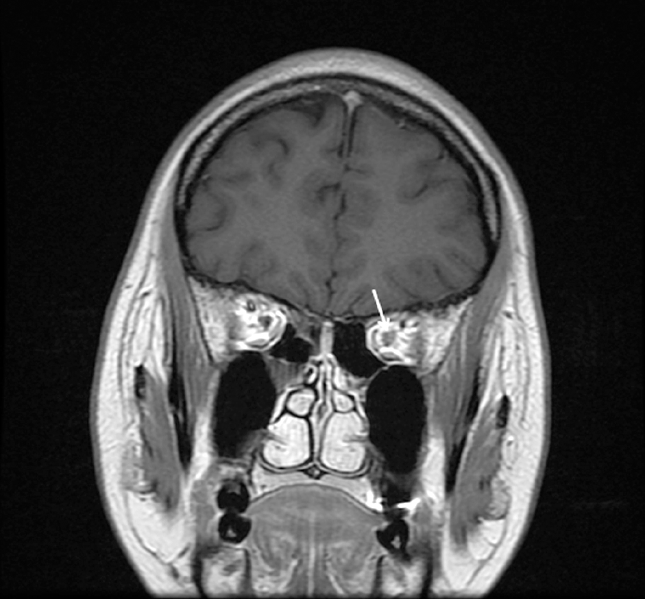
Figure 1.
The picture shows the mild enhancement of left optic nerve (arrow). It suggests that the prominent finding may be left intraorbital optic neuritis.

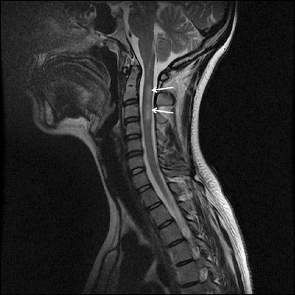
Figure 2.
The picture shows that subtle high signal in cord of C2 and C3 level (arrows) on T2-weighted image. These finding is likely to acute transeverse myelitis in spinal cord of C2-C6 level. It suggests that these findings may be multiple slclerosis.

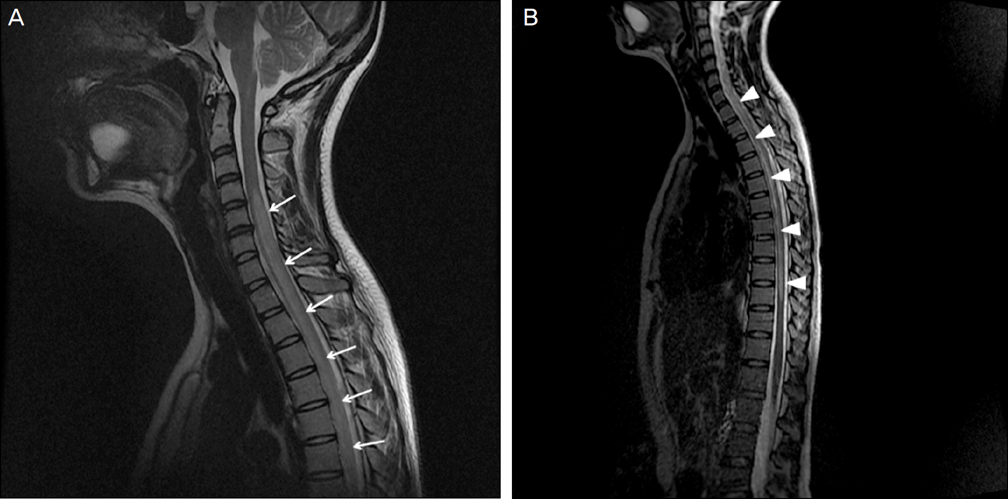
Figure 3.
T2-weighted sequences of spinal cord MR. (A) The picture shows high signal lesion in cord of C2 and C3 level and more extensive lesion in cord of C-spine below C4 and upper T-spine level (arrows). (B) The picture shows the long segment intra-medullary increased signal in spinal cord from C4 to T10 level (arrowheads). It suggests that the finding is likely to neuromyelitis optica.

Table 1.
Proposed diagnostic criteria for optic neuromyelitis2


 PDF
PDF ePub
ePub Citation
 Print
Print


 XML Download