

 PDF
PDF ePub
ePub Citation
 Print
Print


Abstract
Purpose
We investigated the clinical characteristics of ocular trauma in the military for prevention and treatment application.
Methods
We retrospectively surveyed epidemiologic characteristics by investigating the medical records of 790 patients who were hospitalized in the Armed Forces Capital Hospital from January 1, 2001 to December 31, 2010 and investigated the prognostic factors that influenced visual outcome.
Results
Among the 790 patients with ocular trauma, 22.9% of the patients had an open injury and 77.1% had a closed injury. The most common cause of injury was sports-related ocular trauma (39%) and fatigue duty-related trauma (23.4%). The following 8 risk factors were considered poor prognostic factors: open injury, involved posterior segment, operation, initial visual acuity of 0.1 or less, corneal laceration, hyphema, RD, and intraocular foreign body. There was a significant correlation between the probability of poor visual outcome and the number of risk factors (correlation coefficient = -0.468, p < 0.0001).
Go to : 
References
1. Kuhn F, Maisiak R, Mann L. . The ocular Trauma Score (OTS). Ophthalmol Clin North Am. 2002; 15:163–5.

2. Kuhn F, Morris R, Witherspoon CD. Birmingham Eye Trauma Terminology (BETT): terminology and classification of mechan-ical eye injuries. Ophthalmol Clin North Am. 2002; 15:139–43, v.
3. Bae JH, Kim TJ. The prevention of eye injuries in military. J Korean Mil Med Assoc. 2001; 32:48–59.
4. Jung JH, Chung TM, Paik HJ. . A statistical observation of the ocular injuries (I). J Korean Ophthalmol Soc. 1972; 13:239–44.
5. Cho HW, Yoo SH, Ryoo KH. A clinical study of the ocular injuries. J Korean Ophthalmol Soc. 1982; 23:1021–7.
6. Oh TS, Ahn Y, Kim KH. Sports-related ocular injuries. J Korean Ophthalmol Soc. 2001; 42:730–5.
7. Lee YO, Kang DS, Lee KH. A clinical study of the ocular injuries. J Korean Ophthalmol Soc. 1987; 28:395–401.
8. Jang Y, Oh S, Ji NC. A clinical observation of ocular injuries of inpatients. J Korean Ophthalmol Soc. 1993; 34:257–63.
9. Choi JS, Shin KH. Epidemiology of leisure sports-related ocular trauma. J Korean Ophthalmol Soc. 2008; 49:1658–64.
10. Mader TH, Aragones JV, Chandler AC. . Ocular and ocular ad-nexal injuries treated by United States military ophthalmologists during operations desert shield and desert storm. Ophthalmology. 1993; 100:1462–7.
11. Thach AB, Johnson AJ, Carroll RB. . Severe eye injuries in the war in Iraq, 2003-2005. Ophthalmology. 2008; 115:377–82.
12. Chiquet C, Zech JC, Gain P. . Visual outcome and prognostic factors after magnetic extraction of posterior segment foreign bodies in 40 cases. Br J Ophthalmol. 1998; 82:801–6.
13. Esmaeli B, Elner SG, Schork MA, Elner VM. Visual outcome and ocular survival after penetrating trauma: a clinicopathologic study. Ophthalmology. 1995; 102:393–400.
14. Sternberg P Jr, de Juan E Jr, Michels RG, Auer C. Multivariate analysis of prognostic factors in penetrating ocular injuries. Am J Ophthalmol. 1984; 98:467–72.
15. de Juan E, Sternberg P, Michels RG. Penetrating ocular injuries: types of injuries and visual results. Ophthalmology. 1983; 90:1318–22.
16. Gilbert CM, Soong HK, Hirst LW. A two-year prospective study of penetrating ocular trauma at the Wilmer Ophthalmological Institute. Ann Ophthalmol. 1987; 19:104–6.
17. Groessl S, Nanda SK, Mieler WF. Assault-related penetrating ocu-lar injury. Am J Ophthalmol. 1993; 116:26–33.
18. Matthews GP, Das A, Brown S. Visual outcome and ocular survival in patients with retinal detachments secondary to open-or closed- globe injuries. Ophthalmic Surg Lasers. 1998; 29:48–54.
19. Pieramici DJ, MacCumber MW, Humayun MU. . Open-globe injury Update on types of injuries and visual results. Ophthalmology. 1996; 103:1798–803.
20. Dalma-Weiszhausz J, Quiroz-Mercado H, Morales-Canton V. . Vitrectomy for ocular trauma: a question of timing? Eur J Ophthalmol. 1996; 6:460–3.
21. Miyake Y, Ando F. Surgical results of vitrectomy in ocular trauma. Retina. 1983; 3:265–8.
22. Sternberg P Jr, de Juan E Jr, Michels RG. Penetrating ocular in-juries in young patients: initial injuries and visual results. Retina. 1984; 4:5–8.
23. Barr CC. Prognostic factors in corneoscleral lacerations. Arch Ophthalmol. 1983; 101:919–24.
24. Edmund J. The prognosis of perforating eye injuries. Acta Ophthalmol (Copenh). 1968; 46:1165–74.
25. Williams DF, Mieler WF, Abrams GW, Lewis H. Results and prognostic factors in penetrating ocular injuries with retained intra-ocular foreign bodies. Ophthalmology. 1988; 95:911–6.
26. Hutton WL, Fuller DG. Factors influencing final visual results in severely injured eyes. Am J Ophthalmol. 1984; 97:715–22.
27. Russell SR, Olsen KR, Folk JC. Predictors of scleral rupture and the role of vitrectomy in severe blunt ocular trauma. Am J Ophthalmol. 1988; 105:253–7.
28. Punnonen E, Laatikainen L. Prognosis of perforating eye injuries with intraocular foreign bodies. Acta Ophthalmol (Copenh). 1989; 67:483–91.
29. Rahman I, Maino A, Devadason D, Leatherbarrow B. Open globe injuries: factors predictive of poor outcome. Eye. 2006; 20:1336–41.
30. Snell AC Jr. Perforating ocular injuries. Am J Ophthalmol. 1945; 28:263–81.
31. Johnston S. Perforating eye injuries: a five year survey. Trans Ophthalmol Soc U K. 1971; 91:895–921.
32. Cinotti AA, Maltzman BA. Prognosis and treatment of perforating ocular injuries. The John Luhr memorial lecture. Ophthalmic Surg. 1975; 6:54–61.
33. Schmidt GW, Broman AT, Hindman HB, Grant MP. Vision surviv-al after open globe injury predicted by classification and regression tree analysis. Ophthalmology. 2008; 115:202–9.
34. Man CYW, Steel D. Visual outcome after open globe injury: a comparison of two prognostic models— the Ocular Trauma Score and the Classification and Regression Tree. Eye. 2010; 24:84–9.
35. Yoo JH, Lee H, Lee J. . A statistical observation of ocular in-juries and visual predictive value of ocular trauma score. J Korean Ophthalmol Soc. 2011; 52:1024–9.
36. Han SB, Yu HG. Visual outcome after open globe injury and its predictive factors in Korea. J Trauma. 2010; 69:E66–72.
37. Mao CJ, Yan H. Clinical characteristics of mechanical ocular injury and application of ocular trauma score. Zhonghua Yan Ke Za Zhi. 2012; 48:432–5.
38. Unver YB, Kapran Z, Acar N, Altan T. Ocular trauma score in open-globe injuries. J Trauma. 2009; 66:1030–2.
39. Bauza AM, Emami P, Soni N. . A 10-year review of assault-re-lated open-globe injuries at an urban hospital. Graefes Arch Clin Exp Ophthalmol. 2013; 251:653–9.
40. Cho J, Jun BK, Lee YJ, Uhm KB. Factors associated with the poor final visual outcome after traumatic hyphema. Korean J Ophthalmol. 1998; 12:122–9.
Go to :
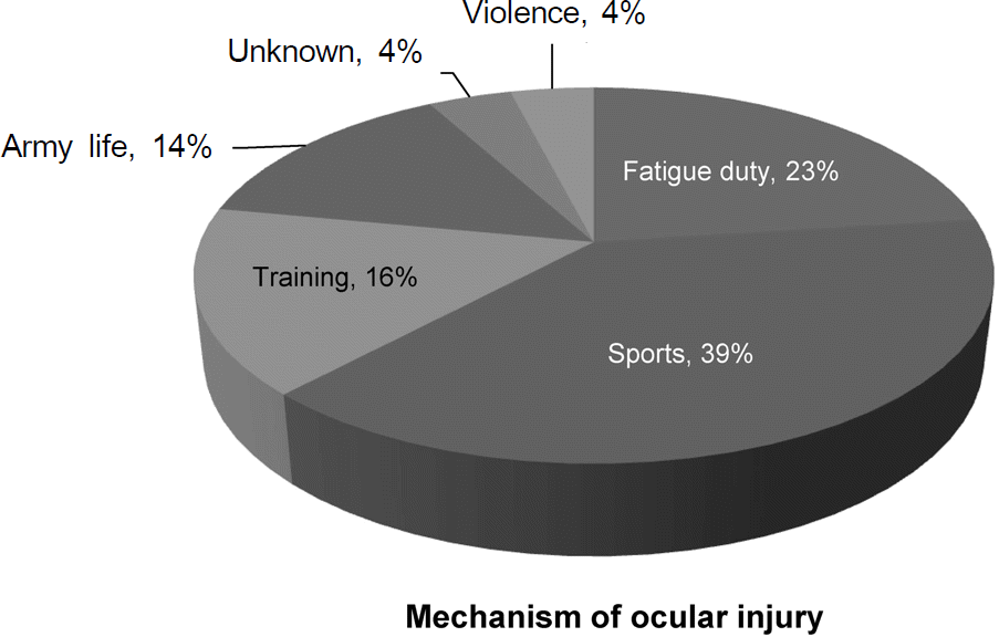
Table 1.
Characteristic of patients
| Characteristic | No. of patients |
|---|---|
| Patients | 790 |
| Sex (male/female) | 789/1 |
| Laterality (OD/OS/OU) | 399/365/26 |
Table 2.
Visual outcomes after management in 531 patients with ocular injury
Table 3.
Univariate analysis of patient characteristics of eight prognostic factors of injured eyes
Table 4.
The relative risks for impossible vision among diagnosis. Patients who had hyphema, corneal laceration and retinal detachment were found to have a greater chance of de-veloping impossible vision than nothing
| p-value | Odds ratio (95% CI) | |
|---|---|---|
| Hyphema | <0.0001 | 2.53 (1.58-4.07) |
| Cornea laceration | 0.001 | 2.32 (1.38-3.89) |
| RD | 0.007 | 3.05 (1.36-6.81) |
 XML Download