

 PDF
PDF ePub
ePub Citation
 Print
Print


Abstract
Purpose
To evaluate the effect of a capsular tension ring (CTR) on the change in postoperative refractive errors and ante-rior chamber depth after cataract surgery in patients with a history of acute primary angle closure (APAC).
Methods
We retrospectively reviewed medical records of 45 patients who underwent cataract surgery with a history of APAC. Forty five eyes were divided into 2 groups based on whether a CTR was implanted (n = 21) or not (n = 24). Spherical equivalent (SE) refractive errors were compared between the 2 groups for 12 months postoperatively. The post-operative anterior chamber depth (ACD) and volume (ACV) were compared between the 2 groups.
Results
SE refractive errors in the CTR group were -0.59 ± 1.08 D at 6 months and -0.61 ± 1.04 D at 12 months postoperatively. In the non-CTR group, -0.64 ± 1.30 D, at 6 months and -0.67 ± 1.19 D at 12 months postoperatively. There was no statistically significant difference between 2 groups (p = 0.89, p = 0.84) and postoperative SE refraction shifted to myopic refractive power in both groups (p < 0.01). The postoperative ACD and ACV also showed no difference between the 2 groups (p = 0.13, p = 0.47).
References
1. Erie JC, Hodge DO, Gray DT. The incidence of primary angle-clo-sure glaucoma in Olmsted County, Minnesota. Arch Ophthalmol. 1997; 115:177–81.

2. Ramli N, Chai SM, Tan GS. . Efficacy of medical therapy in the initial management of acute primary angle closure in Asians. Eye (Lond). 2010; 24:1599–602.
3. Seah SK, Foster PJ, Chew PT. . Incidence of acute primary angle-closure glaucoma in Singapore. An island-wide survey. Arch Ophthalmol. 1997; 115:1436–40.
4. Nongpiur ME, He M, Amerasinghe N. . Lens vault thickness, and position in chinese subjects with angle closure. Ophthalmology. 2011; 118:474–9.
5. Kubota T, Toguri I, Onizuka N. . Phacoemulsification and in-traocular lens implantation for angle closure glaucoma after the re-lief of pupillary block. Ophthalmologica. 2003; 217:325–8.
6. Jacobi PC, Dietlein TS. . Primary phacoemulsification and intraocular lens implantation for acute angle-closure glaucoma. Ophthalmology. 2002; 109:1597–603.
7. Zhuo YH, Wang M, Li Y. . Phacoemulsification treatment of subjects with acute primary angle closure and chronic primary angle-closure glaucoma. J Glaucoma. 2009; 18:646–51.
8. Hayashi K, Hayashi H, Nakao F. . Changes in anterior chamber angle width and depth after intraocular lens implantation in eyes with glaucoma. Ophthalmology. 2000; 107:698–703.
9. Kim SA, Kang JH, Park JI, Lee KH. Difference between post-operative refraction and predictive refraction after cataract oper-ation in patients with coexisting cataract and primary angle-closure glaucoma. J Korean Ophthalmol Soc. 2005; 46:1983–8.
10. Kang SY, Hong S, Won JB. . Inaccuracy of intraocular lens power prediction for cataract surgery in angle-closure glaucoma. Yonsei Med J. 2009; 50:206–10.
11. Sun R, Gimbel HV. In vitro evaluation of the efficacy of the capsu-lar tension ring for managing zonular dialysis in cataract surgery. Ophthalmic Surg Lasers. 1998; 29:502–5.
12. Schild AM, Rosentreter A, Hellmich M. . Effect of a capsular tension ring on refractive outcomes in eyes with high myopia. J Cataract Refract Surg. 2010; 36:2087–93.
13. Kim TW, Kwon JW, Lee JH. Long-term follow-up of the insertion of capsular tension ring in cataract surgery. J Korean Ophthalmol Soc. 2005; 46:1624–9.
14. Cha YD, Kim HJ, Lee DH. New model of capsule measuring ring: The possibility to predict the capsular bag diameter. J Korean Ophthalmol Soc. 2006; 47:221–6.
15. Takimoto M, Hayashi K, Hayashi H. Effect of a capsular tension ring on prevention of intraocular lens decentration and tilt and on anterior capsule contraction after cataract surgery. Jpn J Ophthalmol. 2008; 52:363–7.
16. Nongpiur ME, Sakata LM, Friedman DS. . Novel association of smaller anterior chamber width with angle closure in Singaporeans. Ophthalmology. 2010; 117:1967–73.
17. Wu RY, Nongpiur ME, He MG. . Association of narrow angles with anterior chamber area and volume measured with anterior segment optical coherence tomography. Arch Ophthalmol. 2011; 129:569–74.
18. Wang BS, Narayanaswamy A, Amerasinghe N. . Increased iris thickness and association with primary angle closure glaucoma. Br J Ophthalmol. 2011; 95:46–50.
19. Yoon JY, Hong YJ, Kim CY. Cataract surgery in patients with acute primary angle-closure glaucoma. Korean J Ophthalmol. 2003; 17:122–6.
20. Lam DS, Tham CC, Lai JS, Leung DY. Current approaches to the management of acute primary angle closure. Curr Opin Ophthalmol. 2007; 18:146–51.
21. Kim JE, Park SW. The refractive change after cataract surgery in patients with acute primary angle closure. J Korean Ophthalmol Soc. 2009; 50:1669–73.
22. Hara T, Hara T, Yamada Y. ‘Equator ring’ for maintenance of the completely circular contour of the capsular bag equator after cata-ract removal. Ophhtalmic Surg. 1991; 22:358–9.
23. Cionni RJ, Osher RH. Management of profound zonular dialysis or weakness with a new endocapsular ring designed for scleral fixation. J Cataract Refract Surg. 1998; 24:1299–306.
24. Jacob S, Agarwal A, Agarwal A. . Efficacy of a capsular tension ring for phacoemulsification in eyes with zonular dialysis. J Cataract Refract Surg. 2003; 29:315–21.
25. Park YK, Choi NY, Lee DH, Joo CK. The effect of capsular tension ring on posterior capsular opacity in cataract surgery. J Korean Ophthalmol Soc. 2002; 43:819–22.
26. Lee DH, Shin SC, Joo CK. Effect of a capsular tension ring on in-traocular lens decentration and tilting after cataract surgery. J Cataract Refract Surg. 2002; 28:843–6.
27. Boomer JA, Jackson DW. Effect of the Morcher capsular tension ring on refractive outcome. J Cataract Refract Surg. 2006; 32:1180–3.
Figure 1.
Comparisons of postoperative spherical equivalent between CTR group and Non-CTR group. CTR = capsular tension ring; TR = target refraction; SE = spherical equivalent. * p-values (comparison between TR and postoperative SE in CTR group) < 0.01; † p-values (comparison between TR and postoperative SE in Non-CTR group) < 0.01.
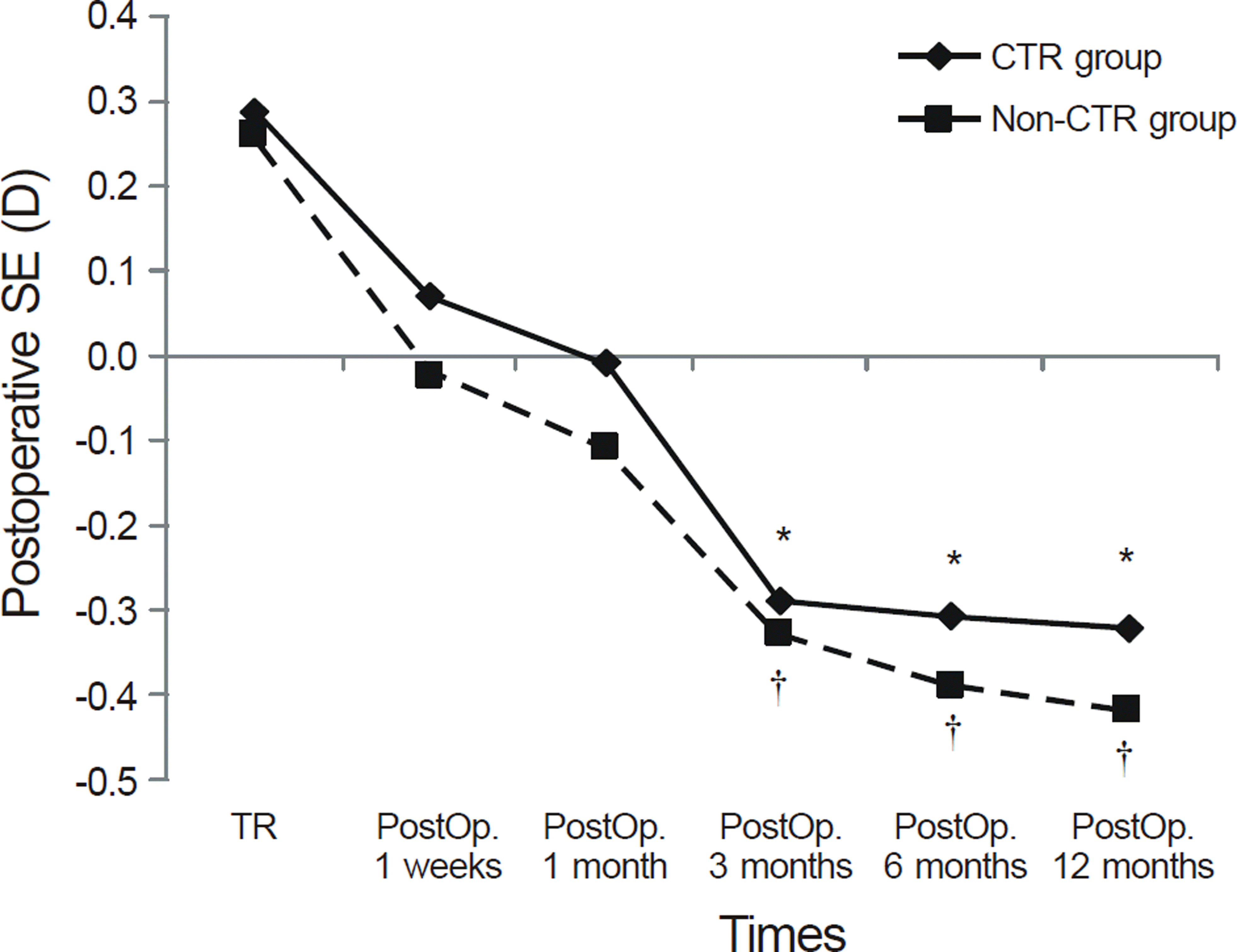
Table 1.
Demographic and clinical data for CTR and Non-CTR group
| CTR group | Non-CTR group | p | |
|---|---|---|---|
| Number of patients | 21 | 24 | |
| Mean age (years) | 65.43 ± 8.74 | 69.79 ± 10.53 | 0.18 |
| Sex (male/female) | 4/17 | 5/19 | 1.00 |
| Axial Length (mm) | 22.69 ±0.80 | 22.01 ± 0.72 | 0.61 |
| Preoperative BCVA (log MAR) | 0.37 ± 0.26* | 0.35 ± 0.28* | 0.83 |
| Postoperative BCVA (log MAR) | 0.15 ± 0.13* | 0.14 ± 0.17* | 0.94 |
| Preoperative IOP (mm Hg) | 18.52 ± 4.99* | 17.17 ± 4.61* | 0.35 |
| Postoperative IOP (mm Hg) | 14.38 ± 2.22* | 13.96 ± 2.99* | 0.59 |
Table 2.
Postoperative spherical equivalent, refractive error and absolute error in CTR group and Non-CTR group
Independent samples t-test; P1, P2, P3. P1 = Comparison of SE between CTR and Non-CTR group; P2 = Comparison of SE-TR between CTR and Non-CTR group; P3 = Comparison of | SE - TR| between CTR and Non-CTR group.
Table 3.
Pre- and post-operative biometric measurements in eyes with CTR group and Non-CTR group
Paired t-test; P1, P2. Independent samples t-test; P3, P4. P1 = Comparison between pre and post operative values in CTR group; P2 = Comparison between pre and postoperative values in Non-CTR group; P3 = Comparison of preoperative values between CTR group and Non-CTR group; P4 = Comparison of postoperative values between CTR group and Non-CTR group. ACD = anterior chamber depth; ACV = anterior chamber volume.
 XML Download