

 PDF
PDF ePub
ePub Citation
 Print
Print


Abstract
Purpose
To investigate the epidemiological and clinical characteristics of industrial ocular trauma for treatment application and prevention.
Methods
A retrospective survey of 207 eyes from 206 patients who visited Gosin University Gospel Hospital from January 1, 1998 to December 31, 2007 was performed. The age, sex, diagnosis, causes, injury site, primary ocular surgery, duration of hospitalization and treatment, and initial and final visual acuities were reviewed using the United States Eye Injury Registry (USEIR) form based on the Birmingham Eye Trauma Terminology (BETT).
Results
The trauma incidence was higher in males (95.65%) in their forties (50.24%). The mean patient age was 41.5 years. Separately counted lesions were presented as a proportion to total injured eyes. The most common diagnosis of industrial ocular traumas was global injuries (124.1%), orbital wall fractures (6.3%), adnexal trauma (5.3%) and optic nerve injuries (3.4%). The most common cause of ocular injuries was flying iron piece (28.67%), and the cornea was the most frequent injured site (69.1%). In 43% of the patients, surgical treatments were performed and the most common surgery was primary closure of the cornea or sclera (82.02%), followed by vitrectomy (30.33%). The average of initial and final visual acuity (log MAR) was 1.2 and 0.93, respectively. In 69.7% of all patients, the final visual acuity was improved or stabilized compared to the initial status.
Conclusions
Flying objects are still the most frequent cause of industrial ocular trauma and in approximately 70% of all patients, the final visual outcome improved or stabilized compared to the initial status. These types of ocular traumas can be significantly reduced by wearing protector shields along with educational safety programs.
References
1. Chung I, Lew HM. A Statistical Observation of Industrial Ocular Injuries in Kangwon-do. J Korean Ophthalmol Soc. 1986; 27:629–38.
2. Shon OO, Kim YJ. An epidemiological Study of occupational abdominal injuries. J Korean Ophthalmol Soc. 1985; 26:531–6.
3. Lee YM, Pak BG. Industrial ocular injuries in Busan area. J Korean Ophthalmol Soc. 1974; 15:335–41.
4. Kim SS, Yoo JM. A clinical study of industrial ocular injuries. J Korean Ophthalmol Soc. 1988; 29:393–403.
5. Kim DI, Kim HK, Hong YJ. A statistical observation of industrial eye injuries. J Korean Ophthalmol Soc. 1982; 23:633–8.
6. Jung JH, Chung TM, Paik HJ, et al. A statistical observation of the ocular injuries(I). J Korean Ophthalmol Soc. 1972; 13:157–61.
7. Kim JY, Kim JW, Lee J. Clinical evaluations of penetration ocular injuries. J Korean Ophthalmol Soc. 1992; 33:919–24.
8. Rhee HC, Chung SM, Rhea SW, Lee WC. Industrial ocular injary in ST. Mary's industrial accident hospital. J Korean Ophthalmol Soc. 1989; 30:995–1001.
9. De Juan E Jr, Sternberg P Jr, Michels RG. Penetrating ocular injuries. Types of injuries and visual results. Ophthalmology. 1983; 90:1318–22.
10. Ahn JW, Moon SH, Lee DH, Lee CY. Factors influencing final abdominal acuiry after penetrating ocular infuries. J Korean Ophthalmol Soc. 1998; 39:2451–8.
11. Pieramici DJ, Au Eong KG, Sternberg P Jr, Marsh MJ. The abdominal significance of a system for classifying mechanical injuries of the eye (globe) in open-globe injuries. J Trauma. 2003; 54:750–4.
12. Jang SG, Lee SJ. Statistical evaluation for perforating ocular injuries. J Korean Ophthalmol Soc. 1988; 29:921–9.
13. Benson WE, Machemer R. Severe perforating injuries treated with pars plana vitrectomy. Am J Ophthalmol. 1976; 81:728–32.

14. Spalding SC, Sternberg P Jr. Controversies in the management of posterior segment ocular trauma. Retina. 1990; 10(Suppl 1):S76–82.
15. Duke-Elder S. System of Ophthalmology. Injuries. London: Henry Kimpton;14:1972. p. 574–6.
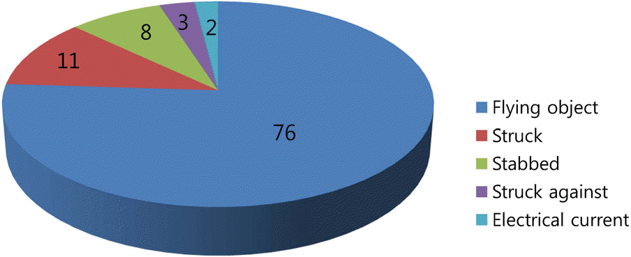
Table 1.
Sex and age distribution of ocular injured patients
Table 2.
The frequency of tissue types experiencing trauma in patients
Table 3.
Specific causes for ocular injuries
Table 4.
Traumatic tissue frequency in patients experiencing open globe trauma
Table 5.
The frequency of treatment types applied to injured eyes
Table 6.
Final visual outcomes by type of injury in open globe injuries
| Final visual acuity* |
Type of Injury |
|||
|---|---|---|---|---|
| Penetrating | IOFB | Rupture | Perforating | |
| Cases (%) | Cases (%) | Cases (%) | Cases (%) | |
| ≥20/40 | 31 (67.0) | 5 (62.5) | 8 (34.2) | 0 |
| 20/50–5/200 | 12 (24.5) | 1 (12.5) | 5 (21.8) | 1 (20.0) |
| <5/200-LP | 3 (7.0) | 2 (25.0) | 7 (31.0) | 3 (60.0) |
| NLP | 1 (1.5) | 0 | 2 (8.3) | 0 |
| Enucleation | 0 | 0 | 1 (4.7) | 1 (20.0) |
Table 7.
Final visual outcomes by grade of injury in open globe injuries
| Final visual acuity* |
Initial visual acuity* |
||||
|---|---|---|---|---|---|
|
≥20/40 |
20/50–20/100 |
19/100–5/200 |
<5/200-LP |
NLP |
|
| Cases (%) | Cases (%) | Cases (%) | Cases (%) | Cases (%) | |
| ≥20/40 | 12 (92.0)) | 7 (77.8) | 9 (64.3) | 16 (38.1) | 0 |
| 20/50–5/200 | 1 (8.0) | 2 (22.2) | 4 (28.6) | 12 (28.6) | 0 |
| <5/200-LP | 0 | 0 | 1 (7.1) | 12 (28.6) | 2 (40.0) |
| NLP | 0 | 0 | 0 | 2 (4.7) | 1 (20.0) |
| Enucleation | 0 | 0 | 0 | 0 | 2 (40.0) |
 XML Download